Patellofemoral Syndrome (PFS)

Patellofemoral Syndrome (PFS), also known as anterior knee pain, is a condition characterized by irritation or damage to the posterior surface of the patella, the kneecap. This syndrome encompasses a spectrum of severity, ranging from mild dysfunction to more serious and chronic issues. The pain originates from the patellofemoral joint or nearby soft tissues. While previously referred to as anterior knee pain, discomfort associated with PFS can be experienced throughout various regions of the knee.

Patellofemoral Syndrome (PFS)

Anatomically, PFS involves the patellofemoral joint, one of the two major joints in the knee. The patella sits within the femoral groove, and if it is displaced, its sharp undersurface may compress against one side of the femoral groove. The patella is connected to the quadriceps muscle tendon and the patellar ligament, playing a crucial role in knee function.

Anatomy

Patellofemoral Syndrome (PFS) involves the patellofemoral joint, which is a crucial component of the knee anatomy. Here’s a more detailed exploration of the relevant anatomical features associated with PFS:

Patellofemoral Joint: The patellofemoral joint is the articulation between the patella (kneecap) and the femur (thigh bone). This joint allows for smooth movement of the patella as the knee flexes and extends.

Patella (Kneecap): The patella is a sesamoid bone embedded in the tendon of the quadriceps muscle. It plays a vital role in the biomechanics of the knee by providing leverage for the quadriceps and protecting the underlying joint surfaces.

Quadriceps Tendon: The quadriceps tendon attaches the quadriceps muscles (rectus femoris, vastus lateralis, vastus medialis, and vastus intermedius) to the patella. This tendon is essential for the extension of the knee.

Patellar Ligament: The patellar ligament continues from the patella and attaches to the tibial tuberosity on the tibia. This ligament is a continuation of the quadriceps tendon and is involved in knee extension.

Femur: The femur is the long bone of the thigh and forms the upper part of the knee joint. The patella glides in the femoral groove during knee movement.

Femoral Groove: The femoral groove is a groove or channel on the femur where the patella moves as the knee flexes and extends. Any deviation in the alignment of the patella within this groove can contribute to PFS.

Quadriceps Vastus Medialis Obliquus (VMO): The VMO is a specific part of the quadriceps muscle located on the inner side of the thigh. It plays a crucial role in stabilizing the patella and preventing lateral displacement.

Meniscus: The menisci (medial and lateral) are crescent-shaped cartilaginous structures located between the femur and tibia. They contribute to shock absorption and load distribution within the knee joint.

Ligaments:

Ligaments around the knee contribute to stability:

Medial Collateral Ligament (MCL): Stabilizes the inner side of the knee.
Lateral Collateral Ligament (LCL): Stabilizes the outer side of the knee.
Anterior Cruciate Ligament (ACL) and Posterior Cruciate Ligament (PCL):

Cruciate ligaments within the joint that control anterior and posterior movement.

Understanding the specific anatomy involved in PFS is crucial for recognizing factors that may contribute to the development of this syndrome. Issues such as malalignment, muscle imbalances, or abnormal patellar tracking can disrupt the normal function of the patellofemoral joint, leading to pain and dysfunction. Proper diagnosis and treatment often involve addressing these anatomical aspects through targeted interventions such as physical therapy, strengthening exercises, and biomechanical assessments.

Symptoms

Patellofemoral syndrome, also known as patellofemoral pain syndrome (PFPS), is a condition characterized by pain in the front of the knee, around the patella (kneecap) and the surrounding area. It is a common cause of knee pain, especially among athletes and individuals engaged in activities that involve repetitive knee motion. The symptoms may include:

• Pain around the kneecap: The primary symptom is usually a dull, aching pain in the front of the knee, especially around or behind the patella. The pain may be aggravated by activities such as climbing stairs, sitting for long periods with the knee bent, or engaging in activities that stress the knee joint.

• Pain during or after activities: Pain may increase during activities that involve bending the knee, such as running, jumping, squatting, or sitting with the knee bent for an extended period.

• Crepitus or grinding sensation: Some individuals with patellofemoral syndrome may experience a grating or grinding sensation (crepitus) when moving the knee.

• Swelling and inflammation: The knee may exhibit signs of swelling or puffiness around the patella.

• Instability or weakness in the knee: Weakness or a feeling of instability in the knee can be present, making it challenging to perform certain activities.

• Pain when sitting for prolonged periods: Individuals with patellofemoral syndrome may experience discomfort when sitting for extended periods, especially with the knee bent, such as during long car rides or at a desk.

It’s important to note that the symptoms of patellofemoral syndrome can vary from person to person, and the severity of symptoms may fluctuate over time. If you suspect you have patellofemoral syndrome or are experiencing persistent knee pain, it is advisable to consult with a healthcare professional, such as a doctor or physical therapist, for an accurate diagnosis and appropriate management plan. Treatment often involves addressing contributing factors, such as muscle imbalances, and implementing exercises and strategies to improve knee function and reduce pain.

Causes

The exact cause of patellofemoral syndrome (PFPS) can be multifactorial, and it often results from a combination of various factors. Some common causes and contributing factors to the development of PFPS include:

• Overuse or Excessive Training: Activities that involve repetitive knee motion, such as running, jumping, or squatting, can lead to overuse of the patellofemoral joint, contributing to the development of PFPS.

• Muscle Imbalances: Weakness or imbalance in the muscles around the knee, particularly the quadriceps (front thigh muscles) and the muscles that make up the hip, can affect the alignment of the patella within its groove. This imbalance may lead to increased stress on the patellofemoral joint.

• Poor Biomechanics: Abnormalities in the way the lower limb moves, such as issues with foot pronation (rolling inward) or supination (rolling outward), can affect the alignment of the patella and contribute to PFPS.

• Flat Feet or Overpronation: Individuals with flat feet or excessive pronation may have altered biomechanics that can lead to increased stress on the patellofemoral joint.

• Malalignment of the Patella: Anatomical factors, such as a misalignment of the patella within its groove, may contribute to PFPS. This can be influenced by factors like the shape and structure of the knee joint.

• Trauma or Injury: Direct trauma to the kneecap or the surrounding structures can contribute to the development of PFPS.

• Tightness or Weakness in the Hamstrings: Imbalances between the quadriceps and hamstring muscles can affect knee joint mechanics, potentially contributing to PFPS.

• Improper Footwear: Wearing shoes that do not provide proper support or that exacerbate biomechanical issues can contribute to PFPS.

• Joint Laxity: Some individuals may have increased joint laxity, which can affect the stability of the patellofemoral joint.

• Changes in Physical Activity: Sudden increases in intensity, duration, or frequency of physical activity can contribute to the development of PFPS.

It’s important to note that these factors alone are not necessarily the only cause, and often, a combination of them may contribute to the development of patellofemoral syndrome. Additionally, individual variations in anatomy and biomechanics can play a role.

Diagnosis

Diagnosing patellofemoral syndrome (PFPS) typically involves a comprehensive evaluation by a healthcare professional. This may include a thorough medical history, a physical examination, and in some cases, imaging studies. Here is an overview of the diagnostic process for PFPS:

• Your doctor or physical therapist will start by asking about your symptoms, including the nature and location of the knee pain, factors that aggravate or alleviate the pain, and any relevant medical history or previous injuries.

• A physical examination will be conducted to assess various aspects of your knee function. This may involve how you walk, assessing the alignment of your lower limbs, and checking for signs of muscle imbalances or weakness. The healthcare provider may also perform specific manoeuvres to reproduce or exacerbate your symptoms.

• Your healthcare provider may palpate (feel) around various structures around the knee, including the patella, to identify areas of tenderness or swelling.

• The range of motion of the knee joint will be assessed, and strength testing of the muscles around the knee, especially the quadriceps and hamstrings, may be conducted.

• Functional tests, such as evaluating your ability to perform specific movements like squatting or climbing stairs, may be used to assess how your knee functions during activities that can aggravate PFPS.

• While the diagnosis of PFPS is often based on clinical evaluation, imaging such as X-rays or MRIs (Magnetic Resonance Imaging) may be ordered to rule out other structural issues and to assess the alignment of the patella and the condition of the surrounding structures.

It’s important to note that the diagnosis of PFPS is often one of exclusion, meaning other knee pain causing factors will be ruled out first.

Treatment

The treatment of patellofemoral syndrome (PFPS) usually involves a combination of conservative measures aimed at reducing pain, improving knee function, and addressing underlying contributing factors. Here are common treatment approaches:

Rest from activities that exacerbate symptoms can help reduce inflammation and pain.

Modifying or temporarily avoiding activities that involve repetitive knee motion, such as running or jumping, may be beneficial.

A physical therapist can design a specific exercise program to strengthen the muscles around the knee, particularly the quadriceps and hip muscles. These can be physiotherapists, sports therapists or personal trainers.

Stretching exercises to improve flexibility in the muscles and tissues around the knee.

Sports Massage or Deep Tissue Massage performed by a qualified therapist who has level 4 or above.

Patellar taping or bracing may be used to help stabilize the patella.

Orthotics and Footwear:

Custom orthotics or supportive footwear may be recommended to address issues related to foot pronation or other biomechanical factors.

Over-the-counter pain medications, such as acetaminophen or nonsteroidal anti-inflammatory drugs (NSAIDs), may be recommended for pain relief.

Ice application can help reduce inflammation. Applying an ice pack to the affected area for about 15-20 minutes at a time can be effective.

Guidance on modifying activities or exercises to reduce stress on the patellofemoral joint.

Evaluation of gait and lower limb biomechanics to identify and address any issues contributing to PFPS.

Maintaining a healthy body weight can help reduce stress on the knee joints.

In some cases, a healthcare provider may recommend corticosteroid injections to reduce inflammation and pain. However, these are typically used judiciously due to potential side effects.

Surgery is rarely the first-line treatment for PFPS. However, in cases where conservative measures are not effective, surgical options such as arthroscopy may be considered. This might involve procedures to address structural issues or correct biomechanical problems.

It’s important to note that the effectiveness of treatment can vary from person to person, and a tailored approach is often necessary. It’s recommended to consult with a healthcare professional, such as an orthopaedic specialist or physical therapist, to determine the most appropriate treatment plan based on individual circumstances. Additionally, early intervention and adherence to recommended exercises and modifications are crucial for successful management of PFPS.

Exercises

Exercise is a key component of the treatment plan for patellofemoral syndrome (PFPS). The goal of exercises is to strengthen the muscles around the knee, improve flexibility, and address any muscle imbalances or weaknesses that may be contributing to the pain. Here are some exercises that are commonly recommended for individuals with PFPS. However, it’s crucial to consult with a healthcare professional or a physical therapist before starting any new exercise program, as the specific exercises should be tailored to individual needs and capabilities.

Quad Sets:

Sit or lie down with your leg extended.

Tighten the muscles on the top of your thigh and push the back of your knee into the surface you’re sitting or lying on.

Hold for a few seconds, then relax.

Repeat for several sets.

Straight Leg Raises:

Lie on your back with one leg straight and the other bent.

Lift the straight leg a few inches off the ground and hold for a few seconds.

Slowly lower the leg back down.

Repeat for several sets on each leg.

Wall Sits:

Stand with your back against a wall and slide down into a sitting position, as if you were sitting in an imaginary chair.

Hold the position for 20-30 seconds.

Stand back up and repeat for several sets.

Step-Ups:

Use a sturdy step or platform.

Step up onto the platform with one foot, then bring the other foot up.

Step back down, leading with the same foot.

Repeat for several sets, alternating legs.

Clamshell Exercise:

Lie on your side with your hips and knees bent at a 90-degree angle.

Keeping your feet together, lift the top knee while keeping the feet together.

Lower the knee back down.

Repeat for several sets on each side.

Lateral Leg Raises:

Lie on your side with the bottom leg bent slightly for support.

Lift the top leg to the side, keeping it straight.

Lower the leg back down.

Repeat for several sets on each side.

Hamstring Stretch:

Sit on the floor with one leg extended and the other bent so that the sole of your foot is against the inner thigh of the extended leg.

Reach toward the toes of the extended leg while keeping your back straight.

Hold the stretch for 20-30 seconds and then switch legs.

Calf Stretch:

Stand facing a wall with your hands on the wall at shoulder height.

Step one foot back and press the heel into the floor.

Hold the stretch for 20-30 seconds and then switch legs.

Remember to start slowly and gradually increase the intensity and duration of exercises. If you experience increased pain or discomfort during any exercise, it’s important to stop and consult with your healthcare provider or physical therapist. Additionally, consistency is key, and performing these exercises regularly as part of a comprehensive rehabilitation program can contribute to improved outcomes for individuals with PFPS.

Prevention

While not all cases of patellofemoral syndrome (PFPS) can be entirely prevented, there are several strategies and lifestyle modifications that can help reduce the risk of developing or exacerbating the condition. Here are some preventive measures:

Strengthening:

Regularly incorporate exercises that focus on strengthening the muscles around the knee, especially the quadriceps, hamstrings, and hip muscles. Strong muscles provide better support to the knee joint.

Flexibility:

Include stretching exercises to maintain flexibility in the muscles and tissues around the knee. This can help prevent muscle imbalances and improve joint function.

Warm-up and Cool Down:

Always warm up before engaging in physical activities or sports. A proper warm-up prepares the muscles and joints for the stresses of exercise. Similarly, cool down with stretching exercises to help prevent stiffness.

Gradual Progression:

Avoid sudden increases in the intensity, duration, or frequency of physical activities. Gradual progression allows your body to adapt to new demands, reducing the risk of overuse injuries like PFPS.

Well Fitted Footwear:

Wear supportive footwear that is appropriate for your activity. Shoes with good arch support and shock absorption can help maintain proper biomechanics and reduce stress on the knee joint.

Maintaining a Healthy Weight:

Excess body weight can contribute to increased stress on the knee joints. Maintaining a healthy weight through a balanced diet and regular exercise can reduce the risk of PFPS.

Biomechanical Assessment:

If you have concerns about your gait or lower limb biomechanics, consider consulting with a healthcare professional or a physical therapist for a biomechanical assessment. Addressing any abnormalities early on can help prevent future issues.

Cross-Training:

Engage in a variety of physical activities to avoid overuse of specific muscle groups or joints. Cross-training can help distribute the workload and reduce the risk of repetitive strain injuries.

Good Technique:

Ensure proper technique when participating in sports or exercises. Improper form can contribute to increased stress on the knee joint and may lead to overuse injuries.

Listen to Your Body:

Pay attention to any signs of pain or discomfort during physical activities. If you experience persistent knee pain, consult with a healthcare professional for evaluation and appropriate management.

It’s important to note that individual factors, such as anatomy and genetics, can also influence the risk of developing PFPS. While these preventive measures can help reduce the risk, there is no one-size-fits-all approach. If you have concerns about your knee health or are experiencing persistent pain, it is advisable to consult with a healthcare professional for personalized guidance and recommendations.

If you believe you may be suffering with PFS then please contact a member of our physiotherapy or sports therapy team.

The Power of Protein: Health and Performance

The Power of Protein: Fueling Your Body for Health and Performance

Protein, often referred to as the building block of life, plays an indispensable role in our overall health and vitality. Beyond the obvious association with muscle growth, protein is a multifaceted nutrient that is involved in numerous essential functions throughout the body. In this article, we’ll explore the power of protein and its critical impact on health, performance, and overall well-being.

What is Protein?

Proteins are complex molecules comprised of amino acids, the fundamental units of life. There are 20 different types of amino acids, and the specific sequence and arrangement of these amino acids in a protein determine its unique function. These functions are diverse and vital, making proteins the workhorses of life.

Building and Repairing Muscles

When most people think of protein, they envision bodybuilders and athletes striving for muscle growth. And they’re not wrong. Protein is crucial for muscle development and repair. When you exercise, you create microscopic tears in your muscle fibers. Protein steps in to repair and strengthen these fibers, resulting in increased muscle mass and strength.

Supporting Enzymes and Hormones

Proteins also serve as enzymes and hormones, which are catalysts for various biochemical reactions in the body. Enzymes are responsible for breaking down food, aiding digestion, and facilitating metabolic processes. Hormones regulate essential functions such as growth, mood, and sleep patterns. Without proteins, these crucial processes wouldn’t function optimally.

Immune Function

Proteins are a cornerstone of the immune system. Antibodies, which defend the body against pathogens, are proteins. A diet rich in protein ensures the body has the raw materials to produce these essential immune defenders.

Transport and Storage

Proteins are involved in transporting vital substances throughout the body. Hemoglobin, for instance, is a protein that carries oxygen from the lungs to tissues, while myoglobin stores oxygen in muscle cells, ensuring it’s available during physical activity.

Maintaining Healthy Skin, Hair, and Nails

Proteins like collagen provide structural support to our skin, hair, and nails. Without adequate protein, you may experience brittle nails, dull hair, and dry skin.

Weight Management

Protein is well-known for its role in weight management. It promotes a feeling of fullness and helps control appetite, which can be instrumental in weight loss and maintenance. It also supports the preservation of lean muscle mass, which is vital for metabolic health.

How Much Protein Do You Need?

The recommended daily intake of protein varies based on factors such as age, sex, activity level, and overall health. On average, a sedentary adult should aim for at least 0.8 grams of protein per kilogram of body weight. However, athletes and individuals looking to build muscle or recover from intense physical activity may require more.

Sources of Protein

Protein is abundant in various foods. Animal sources like lean meats, poultry, fish, and dairy products are complete proteins, meaning they contain all essential amino acids. Plant-based sources like beans, lentils, tofu, and quinoa provide protein too, although they may lack some amino acids found in animal products. Combining plant-based protein sources can create complete proteins for vegetarians and vegans.

In Conclusion

The power of protein cannot be overstated. It is an essential nutrient that influences every aspect of our health and performance. Incorporating an adequate amount of protein into your diet ensures proper muscle development, supports immune function, maintains healthy skin and hair, aids weight management, and provides the body with the tools it needs for optimal function. Whether you’re an athlete striving for peak performance or an individual looking to maintain overall health, protein is a key player in your journey towards well-being and vitality.

If you are interested in how to include more protein in your diet or how much you should be taking on a daily basis, then contact one of our nutritionists and we can provide you with the help and support you need.

The Power of Sports Massage: Performance and Recovery

The Power of Sports Massage: Enhancing Athletic Performance and Recovery

Whether you’re a professional athlete, a weekend warrior, or just someone who enjoys staying active, sports massage can be a game-changer in your fitness journey. This specialized form of massage therapy offers a wide range of benefits that go far beyond relaxation. In this article, we’ll explore the numerous advantages of sports massage and why it’s a crucial component of many athletes’ training and recovery routines.

1. Enhanced Athletic Performance

One of the most significant benefits of sports massage is its ability to boost athletic performance. By targeting specific muscle groups and alleviating tension, sports massage helps improve flexibility and range of motion. This increased flexibility allows athletes to move more freely and efficiently, which is especially vital in sports that require agility and quick movements. As a result, athletes can perform at their best and reduce the risk of injury.

2. Faster Recovery

The demands of intense physical activity can lead to muscle soreness and fatigue. Sports massage helps in the speedy recovery of fatigued muscles by increasing blood circulation and lymphatic flow. This enhanced circulation assists in the removal of waste products and reduces muscle soreness, allowing athletes to bounce back faster and maintain their training consistency.

3. Injury Prevention

Preventing injuries is a top priority for athletes of all levels. Sports massage is an invaluable tool in injury prevention. By identifying and addressing muscle imbalances and areas of tension, massage therapists can reduce the risk of overuse injuries. They can also spot potential issues before they become more severe, helping athletes make necessary adjustments to their training routines.

4. Stress Reduction

Training and competition can be physically and mentally taxing. Sports massage offers a dual benefit by reducing both physical and mental stress. The soothing touch of massage can have a calming effect, helping athletes manage anxiety and stay focused on their goals.

5. Improved Posture and Body Awareness

A well-trained sports massage therapist can identify and address postural issues and imbalances that may be negatively impacting an athlete’s performance. By focusing on these areas, athletes can improve their posture and overall body awareness, which can lead to better movement and reduced strain on the body.

6. Targeted Rehabilitation

In cases where injuries have already occurred, sports massage can be an integral part of the rehabilitation process. Therapists can work alongside healthcare professionals to design a customized massage plan that aids in the healing and recovery of injured muscles and tissues.

7. Enhanced Immune System Function

Regular sports massage can also strengthen the immune system. The reduction of stress and the improvement in circulation and lymphatic flow can help the body better defend against illnesses, keeping athletes in peak condition.

8. Mental Clarity and Focus

Physical well-being and mental health are intricately connected. Sports massage contributes to mental clarity and focus, helping athletes stay motivated and determined to achieve their goals.

In conclusion, sports massage is not just a luxury; it’s a powerful tool that can significantly impact an athlete’s performance, recovery, and overall well-being. Whether you’re a professional athlete or someone who enjoys an active lifestyle, incorporating sports massage into your routine can help you achieve your fitness objectives and enjoy a more balanced, injury-free life. Consult with a qualified sports massage therapist to discover how this specialized therapy can benefit your athletic journey.

Unlocking Employee Wellbeing: A Blueprint for Success

In today’s fast-paced corporate world, where deadlines are relentless and stress is ubiquitous, it’s more critical than ever to prioritize the wellbeing of your most valuable asset – your employees. Happy, healthy employees are not just productive; they’re also more engaged and more likely to stay with your organization in the long run. One company leading the charge in this domain is LiveWell Health, where a team of dedicated experts is transforming corporate wellness through a holistic approach that encompasses on-site massage, reflexology, yoga, mindfulness, mobile injury and rehab clinics, fitness programs, nutrition seminars, and health checks.

On-Site Massage and Reflexology: Stress Relief at Your Fingertips

LiveWell Health recognizes that tension and stress can take a toll on your workforce. That’s why they offer on-site massage and reflexology services. These techniques not only alleviate physical discomfort but also enhance mental clarity and emotional balance. Offering these services in the workplace not only demonstrates a commitment to employee wellbeing but also encourages team members to take breaks and destress during the workday.

Yoga and Mindfulness: A Mind-Body Connection

Yoga and mindfulness practices are essential for creating a mind-body connection that fosters overall wellness. The team at LiveWell Health conducts yoga and mindfulness sessions tailored to the corporate environment. These practices enhance flexibility, reduce stress, and improve mental focus, leading to healthier, happier employees.

Mobile Injury and Rehab Clinics: Preventing and Treating Injuries

To promote physical wellness and prevent workplace injuries, LiveWell Health offers mobile injury and rehab clinics. These clinics provide expert guidance on injury prevention and rehabilitation, helping employees stay physically fit and active.

Fitness at Work: Energizing Your Workforce

LiveWell Health recognizes the importance of regular physical activity. Their fitness programs make it easy for employees to incorporate exercise into their daily routines, boosting energy levels and enhancing productivity.

Nutrition Seminars and Workshops: Fueling Success

A balanced diet is the foundation of overall health. LiveWell Health conducts nutrition seminars and workshops to educate employees about the importance of a healthy diet and how it can improve their performance at work.

Health Checks: A Proactive Approach to Wellbeing

LiveWell Health’s latest offering is health checks that include blood testing for nutrient deficiencies, allergies, and food intolerances. Identifying and addressing these issues can lead to a healthier and more energetic workforce, ultimately reducing absenteeism and enhancing employee satisfaction.

LiveWell Health doesn’t just offer these services in isolation; they tailor their programs to meet the specific needs and objectives of each client. Their team works closely with organizations to design a comprehensive and customized wellbeing strategy, ensuring that every employee is on a journey towards better health and overall satisfaction.

By investing in the wellbeing of your employees through the diverse range of services offered by LiveWell Health, you’re not only creating a healthier workplace but also a more motivated and loyal workforce. The result? Increased productivity, reduced turnover, and a thriving corporate culture. It’s not just about wellness; it’s about unlocking the full potential of your team. LiveWell Health is your partner in this journey, leading the way in redefining corporate wellbeing for the better.

The Winning Edge – Benefits of Event Massage

Introduction: In the competitive world of sports, where athletes push their limits to achieve incredible feats, the role of sports massage therapists has gained significant recognition. Their expertise is more than just relaxation; it’s a crucial component of an athlete’s performance and recovery. Having sports massage therapists on-site during a sporting event to provide your event massage is like having a secret weapon that can provide athletes with the winning edge. In this article, we will delve into the benefits of having sports massage therapists right at the heart of the action.

1. Preparation and Performance Enhancement: Before athletes step onto the field, they need to be in peak physical condition and that is where pre-event massage will help. On-site sports massage therapists are specialists in techniques that can help athletes prepare their bodies for the demands of the game. Through targeted pre-event massages, therapists can stimulate circulation, increase flexibility, and alleviate tension in muscles. This not only enhances performance but also reduces the risk of injuries during their event.

2. Injury Prevention and Rapid Recovery: Athletes are susceptible to injuries due to the repetitive strain and impact on their bodies. On-site sports massage therapists are equipped to address these concerns promptly. By focusing on areas prone to injuries, therapists can identify tightness, knots, or imbalances and use techniques like deep tissue massage, myofascial release, and stretching to alleviate these issues. The immediate attention provided by therapists can prevent minor discomforts from escalating into serious injuries, thereby ensuring athletes stay in the game.

3. Reduced Muscle Fatigue and Improved Circulation: During intense sporting events, athletes’ muscles can become fatigued due to the buildup of lactic acid and other metabolic waste. Sports massage therapists can employ techniques to facilitate the removal of these waste products, promoting better circulation. Improved blood flow delivers oxygen and nutrients to muscles, aiding in their recovery. By mitigating muscle fatigue, athletes can maintain their performance levels throughout the event.

4. Stress and Anxiety Reduction: Competing in high-stakes events can take a toll on an athlete’s mental state. On-site sports massage therapists not only attend to physical concerns but also help athletes manage stress and anxiety. Massage triggers the release of endorphins, which are natural mood enhancers. Athletes can enter competitions with a clear and focused mindset, enabling them to make quick decisions and perform optimally under pressure.

5. Personalized Care and Immediate Feedback: On-site sports massage therapists can provide athletes with individualized attention. They can tailor massage techniques to address specific needs or areas of discomfort, ensuring each athlete receives the care they require. Moreover, therapists can offer immediate feedback regarding an athlete’s muscle condition, flexibility, and overall physical state. This real-time information can guide coaches and athletes in making informed decisions during the event.

6. Enhanced Post-Event Recovery: After the final whistle blows, the importance of post-event recovery cannot be overstated. On-site sports massage therapists continue to play a pivotal role in this phase. Post-event massages help reduce muscle soreness, promote circulation, and expedite the removal of waste products accumulated during the game. Athletes can bounce back quicker, making the recovery process more efficient and effective.

Conclusion: The presence of sports massage therapists on-site at sporting events offers a multitude of benefits that contribute to an athlete’s overall performance and well-being. From preparation and injury prevention to stress reduction and post-event recovery, their expertise serves as a vital component in an athlete’s toolkit for success. As the sports world continues to embrace the importance of holistic care, the role of on-site sports massage therapists remains an essential ingredient in the recipe for victory.

If you are ever in need of a company to help provide your sports massage requirements at your next event, then please contact our team on info@livewellhealth.co.uk or alternatively visit our website and browse our services in more detail by clicking here.


On-Site Chair Massage for Hapag-Lloyd in Liverpool

On-Site Chair Massage for Hapag-Lloyd in Liverpool

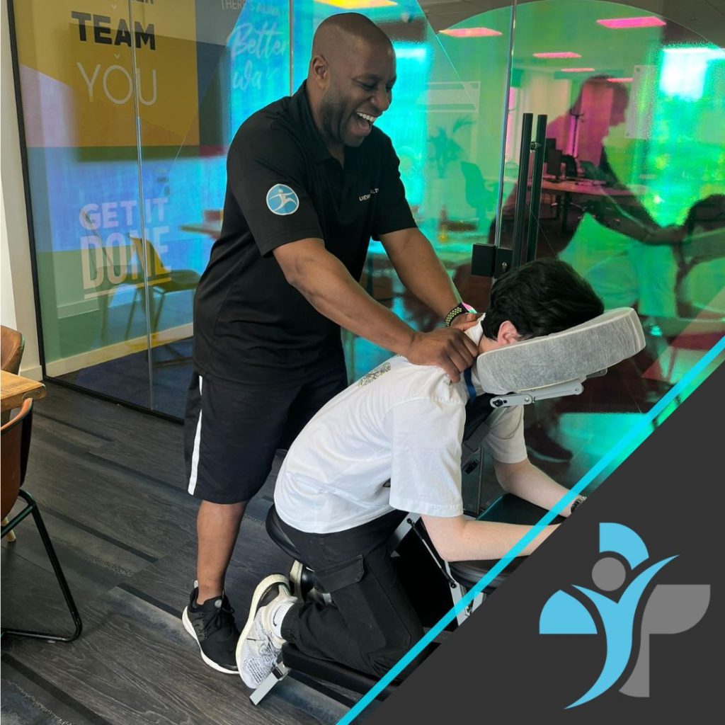
In a remarkable collaboration with Hapag-Lloyd, our team recently had the pleasure of providing on-site chair massages at their Liverpool office. This initiative was a part of Hapag-Lloyd’s ongoing commitment to employee wellbeing, reflecting their dedication to fostering a healthy and productive workplace.

The event took place over the course of a day, with our skilled massage therapists setting up a tranquil space within the office environment. Employees were invited to take a break from their busy schedules and indulge in a relaxing chair massage. The setup was designed to be convenient and comfortable, ensuring that staff could easily participate without disrupting their workday.

Chair massages are known for their numerous benefits, including stress reduction, relief from muscle tension, and improved circulation. Our therapists focused on key areas such as the neck, shoulders, back, and arms, which are often strained during long hours at the desk. Each session lasted about 15 minutes, providing a quick yet effective means for employees to rejuvenate and recharge.

The response from Hapag-Lloyd employees was overwhelmingly positive. Many expressed gratitude for the opportunity to relax and unwind, noting the immediate relief from tension and the overall boost to their mood and productivity. One employee remarked, “It was such a treat to have a massage at work. I feel much more relaxed and ready to tackle my tasks.”

This event underscored the importance of integrating wellness activities into the corporate environment. By prioritising employee health and happiness, Hapag-Lloyd is setting a commendable example for other companies. Our team was delighted to contribute to this effort and looks forward to future collaborations aimed at enhancing employee wellbeing.

In summary, the on-site chair massage event at Hapag-Lloyd’s Liverpool office was a resounding success, highlighting the positive impact of wellness initiatives in the workplace. We are proud to have played a part in this endeavour and are excited about the potential for future partnerships.

If you are interested in Office Massage or any other employee wellbeing initiatives, then please contact our corporate team or visit our contact page.

On-site Chair Massge for Hapag-Lloyd in Liverpool

Hip Arthritis

Osteoarthritis / Arthritis is a degenerative disease where cartilage in the joints of the body get worn down eventually leaving the bone exposed causing endless amount of pain who anyone who suffers with it. Normal or “healthy” cartilage acts as support in the joint, which allows the bone to run smoothly, when damaged it becomes rough causing discomfort for many people.

Anatomy

The hip is a ball and socket joint and is one of the largest joints in the body.  The acetabulum is a part of the pelvis bone which allows the head of the femur to move freely in the socket.

Articular cartilage is a slippery substance which covers the joint, this helps protect and allows free unpainful movement in the ball and socket. Within the hip synovium can be found, this also helps lubricate the joint to allow the hip to function properly.

A person who is suffering with arthritis of the hip will have cartilage that is worn down and will not be producing enough fluid to allow the free movement.  This will lead to the bone rubbing against another bone creating discomfort and pain for an individual.

Hip Arthritis

Symptoms

  • Stiffness around the hip joint
  • Flare ups when walking, running or performing exercise
  • Pain when resting
  • Daily discomfort and ongoing pain in the joint
  • Stiffness in the groin, thigh and glutes when walking
  • Swelling around the hip
  • Grinding sensation in the joint when moving

Causes

  • Arthritis is more common as you get older
  • Family history is a common factor of hip arthritis
  • Individuals who are more overweight often show on going signs of arthritis
  • An injury to the hip and overuse may lead to the long-term effects of arthritis

Diagnosis

A full physical examination will be carried out by a doctor to determine the severity of your condition.

The doctor will carry out special tests specific for the hip to see what movements create the most pain and discomfort. The professional will need to know information such as when the pain first started to occur, how if effects your sleep and how it affects your day-to-day life and activities.

Special tests that are used for the diagnosis of hip arthritis are Trendelenburg test, supine (leg length test) and gait test.

Treatment

Non- surgical treatment that may be effective to help reduce the pain from arthritis are as follows:

  • One of the most successful forms of treatment is to have a deep tissue or sports massage. The reason for this is to lengthen the muscles, relax the muscles around the Hip such as the Glutes etc in order to alleviate some of the pressure in the joint.
  • Ensuring you rest enough when the hip joint is in any pain to reduce irritating the arthritis
  • Preform non-weight baring exercises such as swimming or a gentle cycle to reduce any added pressure onto the hip, this allows the hip to still have good range of motion and keeps the joint functioning
  • Make sure you are getting enough sleep and resting the hip
  • Try eating a healthier balanced diet, this will help with weight loss and reduce the amount of stress placed onto the joints
  • Use over the counter treatment such as deep freeze which may help reduce pain and swelling around the joint

If the pain is ongoing, it is worth speaking to your doctor who may suggest surgery as a last resort. This would mean you would undergo a hip replacement, where you would then follow an 8–12-week rehabilitation programme.

Exercises

These exercises should be performed in sets and reps, it is best to speak to a doctor or physiotherapist to find out the recommended amount suitable for you:

1. Seated Hip Flexion

  • Purpose: Improve hip flexor strength and maintain mobility in the hip joint, which can help alleviate stiffness and pain from arthritis.
  • How to Perform:
    • Sit on a chair with your feet flat on the floor.
    • Lift one knee toward your chest while keeping your back straight.
    • Lower your leg back down slowly and repeat with the other leg.

2. Sit to Stand

  • Purpose: Strengthen the muscles around the hip joint, particularly the quadriceps and glutes, to improve stability and reduce pain from hip arthritis.
  • How to Perform:
    • Sit on a chair with your feet hip-width apart and your hands on your thighs or crossed in front of you.
    • Lean slightly forward and push through your heels to stand up, using your leg muscles rather than your arms.
    • Slowly lower yourself back down to the chair and repeat.

3. Kneeling Hip Flexor Stretch

  • Purpose: Stretch the hip flexors, which can become tight and contribute to pain and reduced mobility in people with hip arthritis.
  • How to Perform:
    • Kneel on one knee with the other foot in front of you, forming a 90-degree angle at both knees.
    • Gently push your hips forward, stretching the front of your hip on the kneeling side.
    • Hold the stretch for 20-30 seconds, then switch legs and repeat.

4. Hip Abduction in Supine Position

  • Purpose: Strengthen the hip abductors, particularly the gluteus medius, which helps support and stabilise the hip joint.
  • How to Perform:
    • Lie on your back with your legs straight and slightly apart.
    • Keeping your leg straight, slowly slide one leg out to the side as far as comfortable, then return it to the starting position.
    • Repeat with the other leg.

5. Hip Extension

  • Purpose: Strengthen the glutes and hamstrings, improving hip stability and reducing pain from arthritis.
  • How to Perform:
    • Stand with your feet hip-width apart and hold onto a sturdy surface for balance.
    • Keeping your leg straight, slowly lift one leg backward, squeezing your glutes.
    • Lower the leg back down slowly and repeat with the other leg.

6. Glute Bridges

  • Purpose: Strengthen the glutes and lower back muscles, providing better support for the hip joint and reducing arthritis symptoms.
  • How to Perform:
    • Lie on your back with your knees bent and feet flat on the floor, hip-width apart.
    • Lift your hips toward the ceiling, squeezing your glutes and keeping your body in a straight line from shoulders to knees.
    • Hold for a few seconds at the top, then slowly lower your hips back down.

7. Body Weight Squat

    • Purpose: Strengthen the quadriceps, hamstrings, and glutes, which helps support and stabilise the hip joint.
    • How to Perform:
      • Stand with your feet shoulder-width apart.
      • Slowly lower your body by bending your knees and hips, keeping your back straight and your weight on your heels.
      • Lower as far as comfortable, then push through your heels to stand back up

Prevention

Prevention of hip arthritis involves making lifestyle changes and taking steps to reduce your risk of developing the condition. Here are some tips for preventing hip arthritis:

  • Maintain a healthy weight: Being overweight or obese puts additional stress on your hip joint, increasing your risk of developing arthritis.
  • Exercise regularly: Regular exercise can help improve your joint health, flexibility, and strength.
  • Avoid overuse injuries: Repeated stress on the hip joint can lead to arthritis over time.
  • Use proper technique: When performing physical activities or exercises, use proper technique
  • Wear appropriate footwear

Avoid sitting for long periods: Sitting for extended periods can increase the pressure on your hips.

Manage other health conditions: Certain health conditions, such as diabetes or rheumatoid arthritis, can increase your risk of developing hip arthritis. Work with your healthcare provider to manage these conditions and reduce your risk.

By making these lifestyle changes and taking steps to reduce your risk of developing hip arthritis, you can help protect your hip joints and maintain good overall health.

If you are suffering with Hip Arthritis and would like to Make a booking or speak with one of our Phsyiotherapists or Sports Therapists then please contact us today.

Hamstring Tear – Grade 1 and 2

Hamstring tear is an injury to a muscle at the back of the thigh, that is caused by a rapid extensive contraction or a violent stretch of the hamstring muscle group, causing high mechanical stress. They account for up to 29% of lower limb injuries in sport, due to the involvement of high speeds such as sprinting and football, however older age can increase the risk due to a loss of flexibility.

Some experiences of hamstring tears involve pain and tenderness, with limited range of motion, where others experience additional symptoms, such as swelling or a ‘popping’ or tearing sensation.

Grade 1 and 2 hamstring tears can heal within 3 to 8 weeks, however can take months to heal depending on the severity of symptoms.

Hamstring Tear – Grade 1 and 2

Anatomy

The hamstrings are a group of three muscles: semitendinosus, semimembranosus, and bicep femoris (long head), that predominantly acts to flex and extend the knee.

Most hamstring tears occur in the central part of the muscle, or where the muscle fibres and tendon fibres intersect (MTJ). Tendons can tear away from a small bone fragment (ischial tuberosity), that typically occur at the proximal origin (top) of the hamstring, however, can occur from the distal origin (bottom), although less common.

Hamstring Tear - Grade 1 and 2

Symptoms

  • Sudden and sharp pain
  • Tenderness
  • A “popping sensation” at the time of injury
  • Swelling and bruising (or discolouration) in the first few hours and days
  • Weakness and a loss of strength in the leg

Causes

Overload of the hamstrings typically the cause of hamstring tears, that occurs when the muscle is stretches beyond its limit or challenged with sudden load, typically during an eccentric contraction. Previous hamstring injuries can increase the risk of developing a tear, particularly if the body is compensating for another limb injury, or if there is muscle weakness and a lack of flexibility.

Diagnosis

Physical examinations are often performed, such as palpation to pinpoint the area of the hamstring tear, however precise location may be difficult to determine without medical imaging.

Other physical examinations involve performing various movement to evaluate strength and range of motion, to determine the severity or the classification (grade 1 to 3) according to the pain and physical limitations.

Neurological examination may be performed to assess the sciatic nerve, to check any entrapment or irritation in healing scar tissue. Imaging such X-rays and MRI may be advised in severe cases, to detect the location of the tear and the extent of the injury, however for grade 1 and 2 tears, only physical examination if typically required.

Treatment

There are 3 phases of treatment to aid recovery and function of the hamstring injury (grade 1 and 2):

Phase 1 (inflammation phase) -Exercises and excessive stretching should be avoided, to control pain and help scar tissue develop. At the phase, crutches may be required to reduce weight bearing and facilitate recovery. You may want to use cold and heat therapy (contrast bathe) as well as gentle massage.

Phase 2 (reparative phase) – Exercises are introduced to try and regain full range of motion. Progressions of more challenging exercises are included in this stage, however, should be performed tolerably and pain-free. Such exercises could include hamstring curls, hip extension (with added resistance band for progression), hamstring wall stretch, and single leg balance.

Phase 3 (remodelling phase) – specific to patients playing sport to allow return to sport, and their level of function. This phase is specific to the patient, where sport or activity-specific drills such as agility are advised to promote tissue remodelling, or drills involving quick movements such as plyometric exercises.

Exercises

  • Hamstring stretch: Sit on the floor with your legs straight out in front of you. Lean forward, reaching your hands towards your toes until you feel a stretch in your hamstrings. Hold for 30 seconds and repeat 3 times.
  • Hamstring curl: Stand facing a wall or chair, with your feet shoulder-width apart. Lift one foot towards your buttocks, keeping your knee bent. Lower your foot and repeat for 10-15 repetitions on each leg.
  • Deadlift: Stand with your feet shoulder-width apart, holding a barbell or dumbbells in front of your thighs. Hinge forward from your hips, keeping your back straight, and lower the weights towards the floor. Return to the starting position and repeat for 10-15 repetitions.
  • Romanian deadlift: Stand with your feet shoulder-width apart, holding a barbell or dumbbells in front of your thighs. Hinge forward from your hips, keeping your back straight, and lower the weights towards the floor. Keep your knees slightly bent and your hips back as you lower the weight. Return to the starting position and repeat for 10-15 repetitions.
  • Glute bridge: Lie on your back with your knees bent and your feet flat on the floor. Lift your hips towards the ceiling, squeezing your glutes at the top. Lower your hips and repeat for 10-15 repetitions. This exercise strengthens the glutes and hamstrings.

Prevention

  • Warm-up properly: Warming up properly before any physical activity is essential to prevent hamstring tears. It increases blood flow to the muscles, making them more pliable and less prone to injury.
  • Stretch regularly: Regular stretching, especially before and after exercise, can help prevent hamstring tears. Incorporate stretching exercises such as hamstring stretches, quadriceps stretches, and hip flexor stretches into your routine.
  • Build strength gradually: Gradually increasing the intensity and duration of your exercise routine can help prevent overloading the hamstrings. Focus on building strength in the hamstrings and surrounding muscles to improve overall stability and reduce your risk of injury.
  • Use proper technique: Using proper technique when performing exercises that involve the hamstrings, such as running and weightlifting, can help prevent hamstring tears. Avoid over-striding, sudden changes of direction, and landing heavily on your heels.
  • Wear appropriate footwear: Wearing appropriate footwear that provides adequate support can help prevent hamstring tears. Choose shoes that fit well and have good shock absorption.

If you are interested in finding out how livewell and our team of physiotherapists, sports therapists and massage therapists can help you in your recovery from your Hamstring tear, then please either book a consultation or contact us today.

Calf Tear

Calf Tear

Calf tears are a relatively common injury that occurs in people of all ages and activity levels. It is estimated that up to 18% of all sports injuries involve the calf muscles, making it one of the most injured areas in the lower leg.

Calf tears are more common in people who engage in sports or activities that involve repetitive or explosive movements of the lower legs, such as running, jumping, or dancing. These activities can put a significant amount of stress on the calf muscles, making them more susceptible to injury.

Calf tears are classified into three grades based on the severity of the injury. Grade 1 tears involve minor damage to the muscle fibers, while grade 2 tears involve a partial tear of the muscle. Grade 3 tears are the most severe and involve a complete tear of the muscle.

While calf tears can be painful and limit mobility, they generally heal well with proper treatment and rehabilitation. It is important to seek medical attention if you suspect a calf tear or if you experience persistent pain or swelling in the calf muscle. With proper care, most people are able to return to their normal activities within a few weeks to a few months.

Anatomy

The calf muscle is made up of two muscles: the gastrocnemius and the soleus. The gastrocnemius is the larger of the two muscles and is responsible for flexing the ankle and knee. The soleus is located underneath the gastrocnemius and is responsible for plantar flexion of the ankle.


Symptoms

The symptoms of a calf tear can vary depending on the severity of the injury. The following are some common symptoms of calf tears grade 1 and 2:

  • Mild to moderate pain in the calf muscle
  • Swelling and tenderness in the affected area
  • Difficulty in walking or standing on the affected leg
  • Stiffness and limited range of motion in the ankle and foot
  • A popping or snapping sensation at the time of injury

Causes

Calf tears can be caused by a sudden or forceful movement, such as pushing off or jumping, which puts excessive strain on the calf muscle. The following are some common causes of calf tears:

  • Overuse or repetitive strain on the calf muscle
  • Sudden movements or changes in direction
  • Inadequate warm-up before exercise or sports activities
  • Poor flexibility or strength in the calf muscles
  • Foot and ankle problems, such as flat feet or ankle instability

Diagnosis

A calf tear can be diagnosed through a physical examination by a healthcare professional. Imaging tests, such as an ultrasound or MRI, may be ordered to confirm the diagnosis and to determine the extent of the injury.

Treatment

The treatment for a calf tear grade 1 or 2 (related article) generally includes the following:

  • Rest: The affected leg should be rested to allow the muscle to heal.
  • Ice: Applying ice to the affected area can help reduce pain and swelling.
  • Compression: Compression with a bandage or brace can help reduce swelling and provide support to the affected area.
  • Elevation: Elevating the affected leg can help reduce swelling and promote healing.
  • Pain medication: Over-the-counter pain medications may be used to help manage pain.

Exercises

After the initial healing period, the following exercises may be prescribed to help improve range of motion and strength in the calf muscle:

  • Calf stretches: Stretching the calf muscle can help improve flexibility and reduce the risk of future injury. Stand facing a wall with your hands on the wall and your feet shoulder-width apart. Step back with your affected leg, keeping your heel on the ground. Lean forward into the wall until you feel a stretch in your calf. Hold for 30 seconds and repeat on the other leg.
  • Calf raises: This exercise helps strengthen the calf muscle. Stand with your feet shoulder-width apart and rise up on your toes, lifting your heels off the ground. Hold for a few seconds and then lower your heels back down. Repeat for 10-15 repetitions.
  • Single leg balance: This exercise helps improve balance and stability in the affected leg. Stand on your affected leg and lift your other leg off the ground. Hold for 30 seconds and repeat on the other leg.

Prevention

To prevent calf tears, it is important to take the following measures:

  • Warm-up: Always warm up before engaging in exercise or sports activities. A proper warm-up can help prepare your muscles for activity and reduce the risk of injury.
  • Stretching: Regular stretching of the calf muscles can help improve flexibility and reduce the risk of tears. Incorporate calf stretches into your warm-up routine and stretch after exercise.
  • Proper footwear: Choose appropriate footwear that provides adequate support and cushioning for your feet and ankles. Replace worn-out shoes regularly.
  • Gradual progression: If you are new to a sport or activity, start slowly and gradually increase the intensity and duration of your workouts. This can help prevent overuse injuries.
  • Strengthening exercises: Regularly performing exercises that strengthen the calf muscles can help prevent tears. Examples include calf raises and resistance band exercises.

By following these preventive measures, you can significantly reduce your risk of calf tears and other lower leg injuries. If you do experience pain or discomfort in your calf muscles, it is important to seek medical attention before continuing with exercise or sports activities.

Wrist Sprain

Wrist sprains occur most often in the athletic or occupational setting but can occur from overuse at work, home, or in any activity of daily living. Injury to the scapholunate ligament is the most common injury and most common form of carpal instability; hyperextension of the wrist is a common mechanism for this type of injury.

An acute wrist sprain is an injury to a ligament often due to an acute traumatic event or chronic repetitive movements. Wrist sprains occur when a ligament is pathologically stretched, twisted, lacerated, or torn.

The inciting event typically involves the sudden application of a force, excessive load-bearing, or twisting injury mechanisms. In most cases, this results from a fall on the outstretched hand with varying structures injured depending on the position of the hand and wrist at the time of injury. In severe cases, there can be a large tear in multiple wrist ligament(s), which can cause instability of the wrist and may require surgical interventions.

Anatomy

A wrist sprain occurs when the ligaments in the wrist are stretched or torn. The ligaments are the bands of tissue that connect the bones in the wrist and help to stabilize the joint.

There are several different ligaments in the wrist, including the radial collateral ligament, ulnar collateral ligament, and the intercarpal ligaments. A sprain can occur in any of these ligaments, but the most common type of wrist sprain is a sprained ligament on the thumb side of the wrist.

Symptoms

The symptoms of a wrist sprain can vary depending on the severity of the injury, but common signs include:

  • Pain: This is the most common symptom of a wrist sprain. The pain is typically located in the affected area and can be sharp, dull, or aching. Pain may be felt when gripping or moving the wrist.
  • Swelling: The wrist may become swollen due to inflammation of the ligaments and the accumulation of fluid in the joint.
  • Bruising: Bruising or discoloration of the skin can occur due to bleeding from the damaged blood vessels.
  • Stiffness: The wrist may feel stiff and difficult to move due to the injury. This is a common symptom of a more severe sprain.
  • Weakness: The affected wrist may feel weak and unstable due to the damage to the ligaments.
  • Instability: The joint may feel loose or unstable, and the wrist may move in an abnormal way.
  • Deformity: In severe cases, there may be a visible deformity of the joint, such as a bend or twist in the wrist.
  • Snapping or popping sensation: Some people may experience a snapping or popping sensation in the joint when they move their wrist.

Causes

There are several common causes of wrist sprains, including:

  • Trauma: A fall or impact to the wrist can cause a sprain. For example, landing on an outstretched hand during a fall can cause a sprain.
  • Overuse: Repetitive motions, such as those involved in sports like gymnastics or tennis, can cause small tears in the ligaments over time, leading to a sprain.
  • Osteoarthritis: This is a degenerative condition that can weaken the ligaments and make them more susceptible to injury.
  • Rheumatoid arthritis: this is an autoimmune disorder that causes inflammation in the joints, leading to damage in the ligaments.
  • Fractures: A fracture can also cause a sprain by damaging the ligaments.
  • Hypermobility: People with hypermobility or ligament laxity are more prone to sprains, as their ligaments are more flexible and can stretch more easily.

Diagnosis

Diagnosis of a wrist sprain typically begins with a physical examination by a healthcare provider. The provider will examine the affected joint for signs of pain, swelling, and tenderness.

They may also move the joint in different directions to assess for range of motion and stability. Imaging tests may also be used to diagnose a wrist sprain. X-rays can help to rule out any fractures and can also show if there is any damage to the bones in the joint. An MRI or CT scan can also be used to confirm a diagnosis and to determine the severity of the injury.

Once a diagnosis of a wrist sprain is made, treatment can begin.

Treatment

Treatment options for a wrist sprain may include:

  • Rest: The affected joint should be rested to allow the ligaments to heal.
  • Ice: Applying ice to the affected area can help to reduce inflammation and pain.
  • Compression: A compression bandage or brace may be used to help reduce swelling and support the joint.
  • Elevation: Keeping the affected joint elevated above the level of the heart can help to reduce swelling.
  • Over-the-counter pain medication: Non-steroidal anti-inflammatory drugs (NSAIDs) such as ibuprofen can help to reduce pain and inflammation.
  • Physical therapy: Physical therapy and rehabilitation exercises may be prescribed to restore range of motion and strength in the joint.
  • Surgery: In severe cases, surgery may be necessary to repair or reconstruct the damaged ligaments.

Recovery time for a wrist sprain can vary depending on the severity of the injury. Mild sprains may take several weeks to heal, while more severe sprains can take several months to heal. It is important to follow the treatment plan and any physical therapy or exercise recommendations provided by a healthcare provider to help ensure a full recovery.

It’s important to note that even after a wrist sprain has healed, it may be more susceptible to future injuries. Therefore, it is important to take steps to prevent future sprains, such as wearing a brace or splint during activities that put the wrist at risk, and doing exercises to strengthen the wrist.

Exercises

The best exercises for a wrist sprain include:

  • Wrist flexion and extension: Gently bend and straighten your wrist to improve range of motion.
  • Wrist pronation and supination: Rotate your wrist to improve strength and flexibility.
  • Wrist circles: Rotate your wrist in both clockwise and counter clockwise directions to improve flexibility.
  • Wrist stretches: Stretch your wrist in different directions to improve flexibility.
  • Grip strengthened exercises: Squeeze a ball or use a hand gripper to improve grip strength.

It is important to consult a doctor or physical therapist before starting any exercise program, particularly if you have a severe sprain. They can advise you on the appropriate exercises for your specific injury and guide you through the healing process.


Prevention

  • Warm up before any physical activity: Take the time to stretch your wrists and hands before participating in any physical activity that requires repetitive wrist motions, such as playing sports or typing.
  • Use proper technique: When participating in sports or other activities that require wrist movements, make sure to use proper technique to avoid putting excessive strain on your wrists.
  • Strengthen your wrists: Regularly performing exercises that strengthen the muscles and tendons in your wrists can help prevent injury. Simple exercises such as wrist curls with light weights or resistance bands can be effective.
  • Take breaks and alternate activities: If you perform repetitive motions with your wrists for extended periods, take frequent breaks to rest and stretch your wrists. Additionally, alternating activities that use different muscle groups can help prevent overuse injuries.
  • Wear appropriate gear: Wearing supportive wrist braces or splints during physical activity can help prevent wrist sprains by providing extra support and stability to the joint.
  • Maintain good posture: Poor posture can put extra strain on your wrists, so make sure to maintain good posture while performing any activity that requires wrist movements.