Nurturing Workplace Happiness: A Comprehensive Guide to Employee Wellbeing
Employee wellbeing is a multifaceted concept that encompasses the physical, mental, and social health of individuals within the workplace. Recognizing the importance of fostering a healthy work environment, companies are increasingly investing in employee wellbeing programs. In this article, we will explore the various dimensions of employee wellbeing and shed light on services, such as office massage, yoga & mindfulness, health checks, exercise at work, nutrition seminars, and in-house mobile physio clinics, that can be brought to the office to enhance the overall wellness of employees. Over 61 million working days are lost on average each and every year through muscular aches and pains and stress related issues. This is where livewell health can help bring a solution that can drastically reduce sickness and absenteeism.
Understanding Employee Wellbeing:
Employee wellbeing goes beyond the absence of illness; it encompasses the overall health, happiness, and engagement of individuals in their work environment. It is influenced by various factors, including physical health, mental and emotional wellness, work-life balance, and the organisational culture.
Bringing Wellness to the Office:
To promote employee wellbeing, companies are increasingly adopting on-site services that cater to the diverse needs of their workforce.
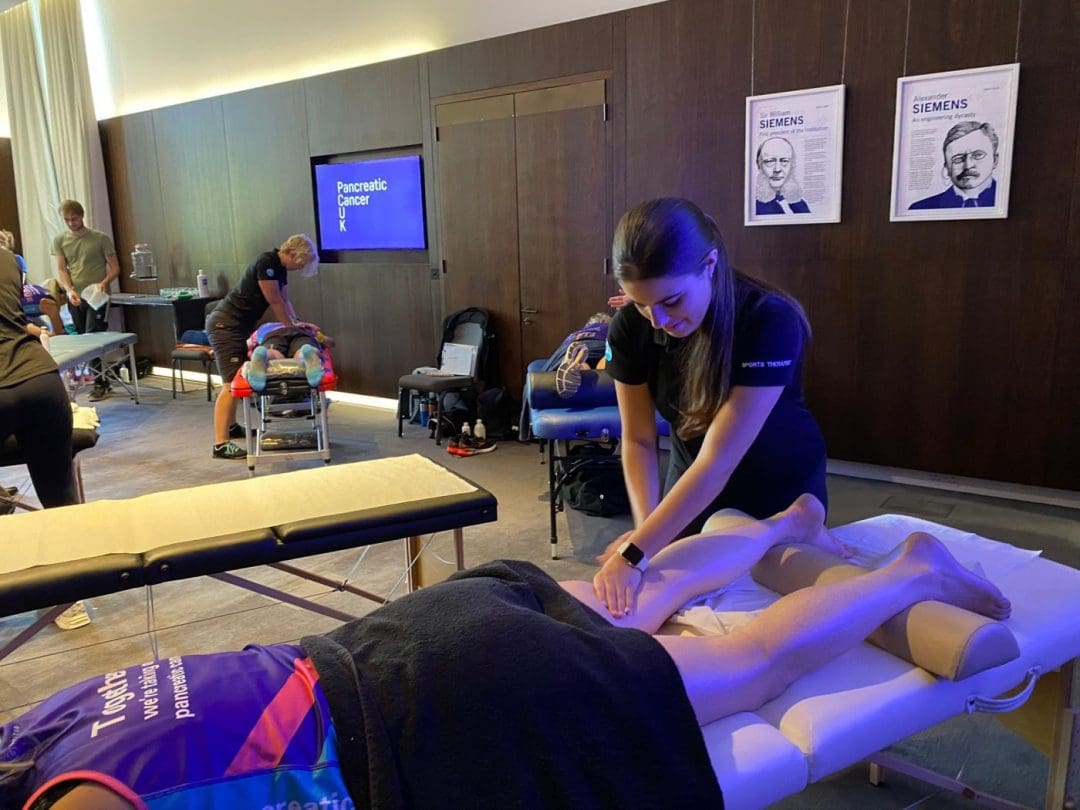
Massage Services:
- On-site massage services, such as those offered by professional practitioners, can significantly contribute to employee wellbeing. Chair massages, within the office, in particular, provide a convenient and accessible way for employees to relax, reduce stress, and alleviate muscle tension during work hours.
Yoga and Mindfulness:
- Incorporating yoga and mindfulness sessions into the workplace can enhance mental and emotional wellbeing. These practices help employees manage stress, improve focus, and foster a positive mindset, contributing to a more resilient and engaged workforce.
Health Checks:
- Regular health checks conducted on-site enable employees to monitor their health status conveniently. These checks can cover a range of parameters, from blood pressure and cholesterol levels to overall fitness assessments, providing employees with valuable insights into their health and motivating them to make positive lifestyle choices.
Exercise at Work:
- Promoting physical activity within the office setting is crucial for maintaining employee health. Simple initiatives, such as standing desks, walking meetings, or on-site fitness classes, encourage employees to incorporate movement into their daily routines.
Nutrition Seminars and Workshops:
- Educating employees about healthy eating habits through on-site nutrition seminars and workshops helps them make informed choices that contribute to overall wellbeing. These sessions can address specific dietary needs and promote a culture of nutritional awareness within the organisation.
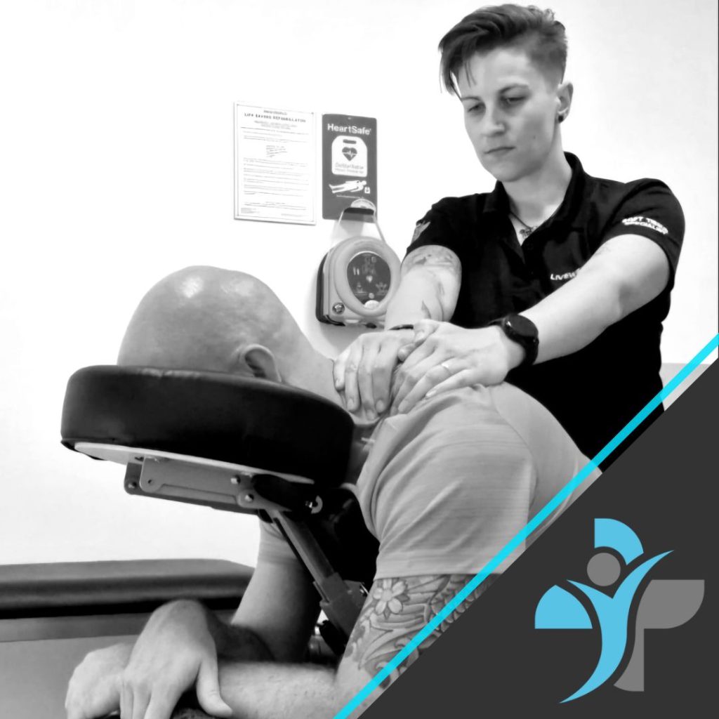
In-House Mobile Physio Clinics:
- On-site physiotherapy clinics provide employees with easy access to musculoskeletal care. Offering services like ergonomic assessments and personalised exercise plans, these clinics contribute to the prevention and management of physical discomfort and injuries.
Subsidised Musculoskeletal Services:
To further support employee wellbeing, companies can extend their commitment by offering subsidised musculoskeletal services beyond the office. Providing access to physiotherapy, fitness, and massage services at home or in external clinics demonstrates a dedication to employee health beyond the workplace.
Conclusion:
Employee wellbeing is a holistic and ongoing commitment that extends beyond traditional benefits. By bringing services like massage, yoga, mindfulness, health checks, exercise at work, nutrition seminars, and in-house mobile physio clinics to the office, companies can create a culture that prioritises the health and happiness of their employees. The ability to extend subsidised musculoskeletal services beyond the workplace underlines a comprehensive approach to employee wellbeing, fostering a positive and supportive work environment.
If you would like to discuss your needs in more detail then please contact us via email or telephone.