Can You Massage Abdominal Adhesions?

Can You Massage Abdominal Adhesions?

Abdominal adhesions are a common yet often overlooked issue that can cause significant discomfort and health problems. These bands of scar tissue form between abdominal organs or between the organs and the abdominal wall, usually as a result of surgery, infection, or inflammation. They can lead to symptoms such as pain, bloating, and even bowel obstruction in severe cases. One question that frequently arises is whether these adhesions can be treated through massage. As soft tissue specialists, LIVEWELL has extensive experience in treating a range of conditions, including abdominal adhesions, using various massage techniques. In this article, we explore whether abdominal adhesions can be effectively managed with massage therapy.

Understanding Abdominal Adhesions

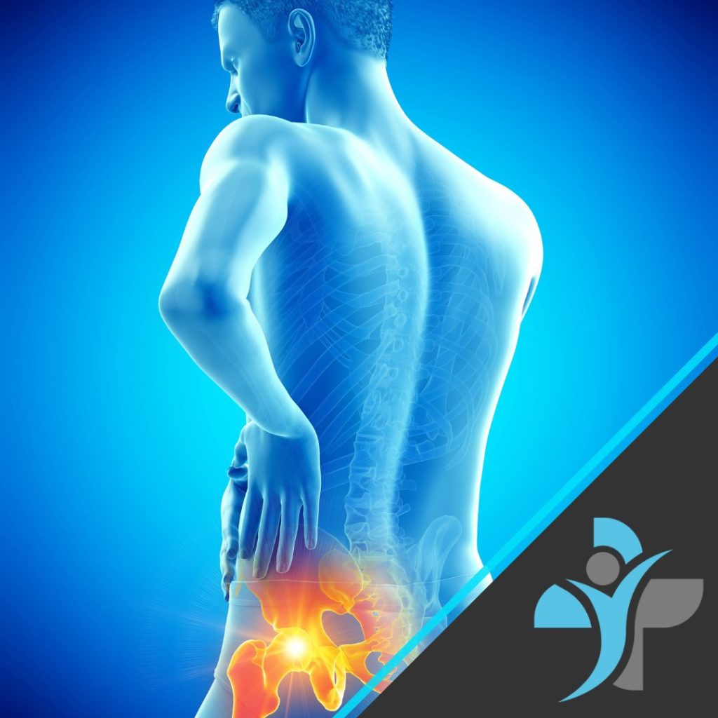
Abdominal adhesions develop when fibrous bands of scar tissue form after abdominal surgery, infection, or inflammation. They can cause organs to stick together or adhere to the abdominal wall, restricting their normal movement. The most common cause of adhesions is surgery, particularly procedures involving the bowel, appendix, or reproductive organs. Even minimally invasive surgeries like laparoscopies can result in adhesions.

In most cases, adhesions are asymptomatic and go unnoticed. However, when they do cause symptoms, they can be debilitating. Chronic pain, digestive issues, and infertility are some of the complications that may arise. In extreme cases, adhesions can lead to bowel obstruction, a life-threatening condition that requires immediate medical attention.

Can Massage Help with Abdominal Adhesions?

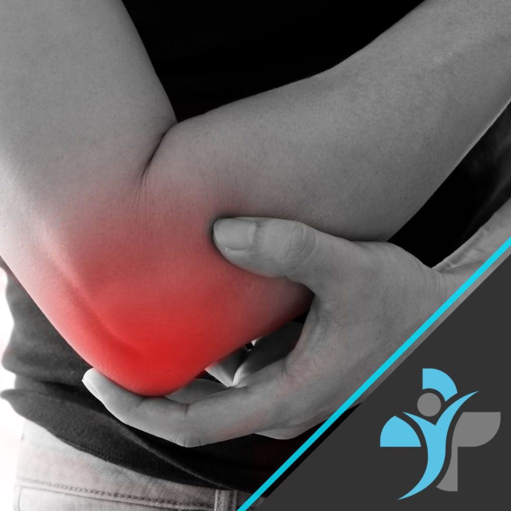
Massage therapy is widely recognised for its ability to treat various musculoskeletal and soft tissue issues, but can it help with abdominal adhesions? The short answer is yes, but with certain caveats. While massage cannot completely eliminate adhesions, it can help to soften and stretch the scar tissue, improving mobility, reducing pain, and alleviating some of the associated symptoms. However, it is crucial that the massage is performed by a skilled therapist with experience in treating adhesions, as improper technique can cause further discomfort or even exacerbate the condition.

How Does Massage Work on Adhesions?

When massage is applied to areas affected by adhesions, it can help to break down some of the fibrous tissue, improving circulation and promoting the healing process. Techniques such as myofascial release, deep tissue massage, and soft tissue mobilisation are particularly effective in treating adhesions. These methods work by gently stretching the scar tissue and surrounding muscles, increasing flexibility and reducing the tension that often accompanies adhesions.

At LIVEWELL, our therapists are trained in a variety of massage techniques, allowing us to tailor treatment to the specific needs of each client. For those suffering from abdominal adhesions, we often use a combination of myofascial release and deep tissue massage to target the affected area. Myofascial release is a gentle technique that focuses on releasing tension in the fascia, the connective tissue that surrounds muscles and organs. By applying sustained pressure to the adhesions, the therapist can help to stretch and soften the scar tissue, improving mobility and reducing pain. Deep tissue massage, on the other hand, works to relieve deeper layers of muscle and connective tissue, addressing the root cause of the discomfort.

The Importance of Professional Treatment

While massage can be an effective treatment for abdominal adhesions, it is essential that it is performed by a qualified professional. The abdomen is a sensitive area, and improper technique can lead to injury or aggravate existing adhesions. At LIVEWELL, our therapists are among the most highly qualified in the UK, with extensive training in a wide range of massage modalities, including Swedish Massage, deep tissue, sports massage, lymphatic drainage, and pregnancy massage. This breadth of expertise ensures that we can provide safe and effective treatment for a wide range of conditions, including abdominal adhesions.

Our team of soft tissue specialists is also well-versed in the anatomy and physiology of the abdomen, enabling us to understand the complexities of adhesions and how best to treat them. Whether you require treatment in one of our clinics or prefer the convenience of a mobile service, our nationwide coverage ensures that we can reach you no matter where you are located in the UK.

The Benefits of Massage for Abdominal Adhesions

When performed correctly, massage therapy can offer several benefits for those suffering from abdominal adhesions. These include:

  1. Pain Relief: Adhesions can cause chronic pain by pulling on surrounding tissues and restricting movement. Massage helps to release this tension, reducing pain and discomfort.
  2. Improved Mobility: By softening the scar tissue, massage can help to restore the normal movement of organs and muscles, improving overall mobility.
  3. Enhanced Circulation: Massage promotes better blood flow to the affected area, aiding in the healing process and reducing inflammation.
  4. Digestive Health: Abdominal adhesions can interfere with digestion, leading to bloating and other gastrointestinal issues. Massage can help to alleviate these symptoms by promoting better movement of the abdominal organs.
  5. Prevention of Further Complications: Regular massage therapy can help to maintain the flexibility of the tissues, reducing the risk of further complications such as bowel obstruction.

A Holistic Approach to Treatment

At LIVEWELL, we believe in taking a holistic approach to treatment. While massage can be highly effective in managing abdominal adhesions, it is often most beneficial when combined with other therapies and lifestyle changes. For example, we may recommend a program of gentle stretching exercises to complement the massage, helping to maintain flexibility and prevent the recurrence of adhesions. We may also suggest dietary adjustments to reduce inflammation and promote better digestive health.

For clients who have undergone surgery, post-operative care is crucial in preventing the formation of adhesions. Our therapists are trained in post-surgical massage techniques that can help to minimise scar tissue development and promote faster healing. Additionally, we offer lymphatic drainage massage, which can reduce swelling and improve circulation, further aiding in the recovery process.

When to Seek Professional Help

While massage can be an effective treatment for abdominal adhesions, it is not always appropriate in every case. If you are experiencing severe pain, digestive issues, or symptoms of bowel obstruction, it is essential to seek medical advice before undergoing massage therapy. In some cases, surgery may be necessary to address the adhesions, and massage should only be considered as part of a broader treatment plan.

It is also important to note that massage is not a quick fix. Depending on the severity of the adhesions, it may take several sessions to achieve noticeable results. At LIVEWELL, we work closely with our clients to develop a personalised treatment plan that meets their needs and goals. We take the time to understand each client’s unique situation and provide ongoing support and advice to help them achieve the best possible outcome.

The difference between our therapists and other companies?

What sets LIVEWELL apart is our commitment to excellence and our dedication to client care. We are proud to be the UK’s leading provider of mobile and clinic-based massage therapy, offering a level of expertise and professionalism that is unmatched in the industry. Our therapists are not only highly skilled but also passionate about helping clients achieve their health and wellness goals. Whether you are dealing with abdominal adhesions or any other soft tissue issue, you can trust that you are in the best possible hands with LIVEWELL.

Our extensive range of qualifications means that we can offer a wide variety of treatments, tailored to the individual needs of our clients. From Swedish massage to sports massage, deep tissue to lymphatic drainage, we have the skills and knowledge to address a wide range of conditions. Our nationwide coverage also means that we can provide our services wherever you are, whether you prefer to visit one of our clinics or enjoy the convenience of a mobile service.

Conclusion

Abdominal adhesions can be a challenging and painful condition, but with the right treatment, they can be managed effectively. Massage therapy, when performed by a qualified professional, can help to soften and stretch scar tissue, improve mobility, and reduce pain. At LIVEWELL, our team of soft tissue specialists is uniquely equipped to provide this type of treatment, offering both clinic and mobile services across the UK. With our extensive range of qualifications and our commitment to client care, we are proud to be the UK’s leading provider of massage therapy.

If you are struggling with abdominal adhesions or any other soft tissue condition, we invite you to experience the LIVEWELL difference. Contact us today to find out how we can help you achieve better health and well-being through our expert massage services. Alternatively use our booking system to book directly.

What is Tennis Elbow?

What is Tennis Elbow?

Tennis elbow, officially termed lateral epicondylitis, affects more than just athletes. This prevalent condition involves pain and inflammation in the tendons that link the forearm muscles to the outer part of the elbow.

Repetitive actions such as gripping, twisting, and lifting can often cause this ailment. Activities like going to your local gym, painting, carpentry, and even typing may trigger these motions, leading to small tears in the tendons. Consequently, this results in inflammation and discomfort on the elbow’s exterior, which can intensify with continued use. Symptoms may also include forearm weakness, reduced grip strength, elbow stiffness, and pain extending down the arm.

What is tennis elbow

To diagnose tennis elbow, a physical examination is typically performed alongside a review of your medical history and activities. Imaging tests might be used to exclude other potential conditions but in the main a sports therapist, physiotherapist or doctor can accurately diagnose this condition.

Treatment aims to alleviate pain, reduce inflammation, and facilitate healing. This can involve rest, ice application, specific physical therapy exercises, bracing, over-the-counter pain relievers, or sometimes corticosteroid injections. In general we see huge improvements in the condition through a well strategised and dianamic recoveyr and rehabilitation regime.

Preventing tennis elbow is crucial, particularly if your job or hobbies involves repetitive arm movements. It is important to warm up before engaging in such activities, maintain proper form, take regular breaks, strengthen forearm muscles, and use ergonomic tools when necessary.

If you think you might have tennis elbow, it’s important to consult a healthcare provider for a precise diagnosis and tailored treatment plan. With prompt intervention and appropriate care, you can effectively manage the condition and resume your regular activities. If you would like to read more about the condition, we have written an article that goes into much more detail which can be found here.

Alternatively if you would like to book one of our sports therapists or physiotherapists to conduct a thorough in person review, treatment and then provide you with the exercises to rehabilitate this condition then please either make a booking through our online booking system or contact us directly.

Nerve Impingement

Nerve impingement, also known as nerve compression or pinched nerve, is a condition in which a nerve is compressed or constricted, leading to pain, weakness, numbness, or tingling sensations in the affected area. This can occur in various parts of the body, including the neck, back, arms, and legs.
The pathology of nerve impingement involves pressure or constriction on the nerve, which can be caused by a variety of factors, such as injury, repetitive motions, poor posture, or degenerative conditions like arthritis. This pressure can lead to irritation and inflammation of the nerve, which can cause pain and other symptoms.
The occurrence of nerve impingement is quite common, especially as people age and degenerative changes occur in the spine. It can also be seen in people who perform repetitive motions or maintain poor posture for extended periods of time. Additionally, nerve impingement can result from injuries, such as whiplash or a herniated disc, or from conditions such as carpal tunnel syndrome or sciatica.

Anatomy

The human nervous system is comprised of a complex network of nerves that run throughout the body, transmitting signals between the brain and various tissues and organs. Nerves are made up of axons, which carry electrical signals, and are surrounded by a layer of protective tissue called myelin. Nerves can be affected by a variety of factors, including compression, entrapment, inflammation, and injury.


Symptoms

The symptoms of nerve impingement can vary depending on the location and severity of the impingement. Common symptoms may include pain, numbness, tingling, burning, or a feeling of pins and needles. In severe cases, nerve impingement can lead to muscle weakness, loss of sensation, and difficulty with movement.

Causes

Nerve impingement can be caused by a variety of factors, including physical trauma, repetitive motions, poor posture, and underlying medical conditions such as arthritis, diabetes, and nerve disorders. Other common causes of nerve impingement include spinal stenosis, herniated discs, and degenerative conditions such as spinal cord injuries.

People who are at a higher risk of developing nerve impingement include:

  • Individuals with certain medical conditions such as diabetes, arthritis, or other conditions that can cause nerve damage or inflammation.
  • Athletes and physically active individuals who engage in repetitive motions, such as running, cycling, or weightlifting, which can put stress on the nerves.
  • People who have a sedentary lifestyle and spend long hours sitting or working in positions that can put pressure on the nerves, such as office workers or truck drivers.
  • Individuals with a family history of nerve impingement, as this may suggest a genetic predisposition to the condition.
  • People who have suffered a previous injury, such as a fracture or dislocation, that may have damaged the nerves.

Diagnosis

The diagnosis of nerve impingement is typically based on a review of medical history, a physical examination, and diagnostic imaging tests such as MRI or CT scans. Nerve conduction studies and electromyography (EMG) tests can also be used to help diagnose nerve impingement and determine the severity of the injury.

It is important to note that while anyone can develop nerve impingement, early detection and proper treatment can greatly reduce the risk of developing long-term complications and help ensure a full recovery.

Treatment

Here are some of the treatment options for nerve impingement:

Rest: Resting the affected area can help reduce inflammation and give the nerve time to heal. You may need to avoid activities that aggravate your symptoms, such as heavy lifting, repetitive motions, or prolonged sitting or standing.

Physical therapy: Physical therapy can help improve your strength, flexibility, and posture, which can relieve pressure on the affected nerve. Your physical therapist may recommend exercises, stretches, or other techniques to help alleviate your symptoms.

Medications: Non-steroidal anti-inflammatory drugs (NSAIDs) like ibuprofen or naproxen can help reduce inflammation and relieve pain associated with nerve impingement. In some cases, corticosteroids may be prescribed to reduce inflammation and alleviate symptoms.

Injections: In some cases, injections of corticosteroids or other medications may be given directly into the affected area to reduce inflammation and alleviate symptoms.

Surgery: If conservative treatments are not effective, or if nerve impingement is causing significant weakness or loss of function, surgery may be recommended. The type of surgery will depend on the location and severity of the compression and may involve removing bone or tissue to relieve pressure on the affected nerve.

Lifestyle changes: Making lifestyle changes can help prevent or alleviate nerve impingement. This may include maintaining good posture, staying active, avoiding repetitive motions, and avoiding activities that put excessive strain on your nerves.

Exercises

Exercises to help relieve nerve impingement and prevent further damage may include:

  • Stretching: Gentle stretching exercises can help relieve pressure on the nerves and improve flexibility in the affected area.
  • Strengthening exercises: Targeting the muscles surrounding the affected nerve can help relieve pressure and prevent further damage.
  • Posture correction: Paying attention to posture and correcting any imbalances can help reduce stress on the nerves.
  • Aerobic exercises: Engaging in low-impact aerobic activities, such as swimming or walking, can help improve blood flow to the affected area and promote healing.
  • Yoga or Pilates: Gentle yoga or Pilates can help improve flexibility and strengthen the muscles surrounding the affected nerve.

It is important to consult with a doctor or physical therapist before beginning any exercise program, as they can help determine which exercises are best for your individual needs and ensure proper form and technique.

Prevention

Maintain Correct Posture:

Be mindful of your posture, especially when sitting or standing for long periods. Maintain a neutral spine position to reduce unnecessary stress on the spine and nerves.

Ergonomics:

Ensure that your workspace, whether at a desk or using electronic devices, is ergonomically designed to support good posture. Use supportive chairs, maintain eye-level screens, and take regular breaks to avoid prolonged periods of the same position.

Regular Exercise:

Engage in regular physical activity to strengthen the muscles that support the spine. Focus on exercises that promote core strength and flexibility, as a strong and flexible spine is less prone to nerve compression.

Correct Lifting Techniques:

When lifting objects, use proper lifting techniques to avoid putting excessive strain on the spine. Bend at the knees, keep the back straight, and lift with the legs rather than the back.

Weight Management:

Maintain a healthy weight to reduce the load on the spine. Excess body weight can contribute to conditions like herniated discs and spinal stenosis, increasing the risk of nerve impingement.

Stay Hydrated:

Hydration is essential for maintaining the elasticity of spinal discs. Dehydration can contribute to disc degeneration, which may increase the risk of nerve compression.

Avoid Repetitive Stress:

Be mindful of repetitive movements or activities that can strain specific nerves. If your work or hobbies involve repetitive motions, take breaks and incorporate stretching exercises to reduce the risk of overuse injuries.

Regular Stretching:

Include regular stretching exercises in your routine to maintain flexibility and prevent muscle imbalances. Focus on stretches that target the muscles around the spine, including the neck, back, and hips.

Quit Smoking:

Smoking has been associated with increased disc degeneration and reduced blood flow to spinal structures. Quitting smoking can contribute to overall spine health.

Proper Nutrition:

Maintain a balanced diet rich in essential nutrients, as proper nutrition supports the health of spinal structures. Calcium and vitamin D are particularly important for bone health.

Regular Check-ups:

Schedule regular check-ups with your healthcare provider to monitor your spine health. Early detection and management of spinal conditions can help prevent the progression to nerve impingement.

Hip Arthritis

Osteoarthritis / Arthritis is a degenerative disease where cartilage in the joints of the body get worn down eventually leaving the bone exposed causing endless amount of pain who anyone who suffers with it. Normal or “healthy” cartilage acts as support in the joint, which allows the bone to run smoothly, when damaged it becomes rough causing discomfort for many people.

Anatomy

The hip is a ball and socket joint and is one of the largest joints in the body.  The acetabulum is a part of the pelvis bone which allows the head of the femur to move freely in the socket.

Articular cartilage is a slippery substance which covers the joint, this helps protect and allows free unpainful movement in the ball and socket. Within the hip synovium can be found, this also helps lubricate the joint to allow the hip to function properly.

A person who is suffering with arthritis of the hip will have cartilage that is worn down and will not be producing enough fluid to allow the free movement.  This will lead to the bone rubbing against another bone creating discomfort and pain for an individual.

Hip Arthritis

Symptoms

  • Stiffness around the hip joint
  • Flare ups when walking, running or performing exercise
  • Pain when resting
  • Daily discomfort and ongoing pain in the joint
  • Stiffness in the groin, thigh and glutes when walking
  • Swelling around the hip
  • Grinding sensation in the joint when moving

Causes

  • Arthritis is more common as you get older
  • Family history is a common factor of hip arthritis
  • Individuals who are more overweight often show on going signs of arthritis
  • An injury to the hip and overuse may lead to the long-term effects of arthritis

Diagnosis

A full physical examination will be carried out by a doctor to determine the severity of your condition.

The doctor will carry out special tests specific for the hip to see what movements create the most pain and discomfort. The professional will need to know information such as when the pain first started to occur, how if effects your sleep and how it affects your day-to-day life and activities.

Special tests that are used for the diagnosis of hip arthritis are Trendelenburg test, supine (leg length test) and gait test.

Treatment

Non- surgical treatment that may be effective to help reduce the pain from arthritis are as follows:

  • One of the most successful forms of treatment is to have a deep tissue or sports massage. The reason for this is to lengthen the muscles, relax the muscles around the Hip such as the Glutes etc in order to alleviate some of the pressure in the joint.
  • Ensuring you rest enough when the hip joint is in any pain to reduce irritating the arthritis
  • Preform non-weight baring exercises such as swimming or a gentle cycle to reduce any added pressure onto the hip, this allows the hip to still have good range of motion and keeps the joint functioning
  • Make sure you are getting enough sleep and resting the hip
  • Try eating a healthier balanced diet, this will help with weight loss and reduce the amount of stress placed onto the joints
  • Use over the counter treatment such as deep freeze which may help reduce pain and swelling around the joint

If the pain is ongoing, it is worth speaking to your doctor who may suggest surgery as a last resort. This would mean you would undergo a hip replacement, where you would then follow an 8–12-week rehabilitation programme.

Exercises

These exercises should be performed in sets and reps, it is best to speak to a doctor or physiotherapist to find out the recommended amount suitable for you:

1. Seated Hip Flexion

  • Purpose: Improve hip flexor strength and maintain mobility in the hip joint, which can help alleviate stiffness and pain from arthritis.
  • How to Perform:
    • Sit on a chair with your feet flat on the floor.
    • Lift one knee toward your chest while keeping your back straight.
    • Lower your leg back down slowly and repeat with the other leg.

2. Sit to Stand

  • Purpose: Strengthen the muscles around the hip joint, particularly the quadriceps and glutes, to improve stability and reduce pain from hip arthritis.
  • How to Perform:
    • Sit on a chair with your feet hip-width apart and your hands on your thighs or crossed in front of you.
    • Lean slightly forward and push through your heels to stand up, using your leg muscles rather than your arms.
    • Slowly lower yourself back down to the chair and repeat.

3. Kneeling Hip Flexor Stretch

  • Purpose: Stretch the hip flexors, which can become tight and contribute to pain and reduced mobility in people with hip arthritis.
  • How to Perform:
    • Kneel on one knee with the other foot in front of you, forming a 90-degree angle at both knees.
    • Gently push your hips forward, stretching the front of your hip on the kneeling side.
    • Hold the stretch for 20-30 seconds, then switch legs and repeat.

4. Hip Abduction in Supine Position

  • Purpose: Strengthen the hip abductors, particularly the gluteus medius, which helps support and stabilise the hip joint.
  • How to Perform:
    • Lie on your back with your legs straight and slightly apart.
    • Keeping your leg straight, slowly slide one leg out to the side as far as comfortable, then return it to the starting position.
    • Repeat with the other leg.

5. Hip Extension

  • Purpose: Strengthen the glutes and hamstrings, improving hip stability and reducing pain from arthritis.
  • How to Perform:
    • Stand with your feet hip-width apart and hold onto a sturdy surface for balance.
    • Keeping your leg straight, slowly lift one leg backward, squeezing your glutes.
    • Lower the leg back down slowly and repeat with the other leg.

6. Glute Bridges

  • Purpose: Strengthen the glutes and lower back muscles, providing better support for the hip joint and reducing arthritis symptoms.
  • How to Perform:
    • Lie on your back with your knees bent and feet flat on the floor, hip-width apart.
    • Lift your hips toward the ceiling, squeezing your glutes and keeping your body in a straight line from shoulders to knees.
    • Hold for a few seconds at the top, then slowly lower your hips back down.

7. Body Weight Squat

    • Purpose: Strengthen the quadriceps, hamstrings, and glutes, which helps support and stabilise the hip joint.
    • How to Perform:
      • Stand with your feet shoulder-width apart.
      • Slowly lower your body by bending your knees and hips, keeping your back straight and your weight on your heels.
      • Lower as far as comfortable, then push through your heels to stand back up

Prevention

Prevention of hip arthritis involves making lifestyle changes and taking steps to reduce your risk of developing the condition. Here are some tips for preventing hip arthritis:

  • Maintain a healthy weight: Being overweight or obese puts additional stress on your hip joint, increasing your risk of developing arthritis.
  • Exercise regularly: Regular exercise can help improve your joint health, flexibility, and strength.
  • Avoid overuse injuries: Repeated stress on the hip joint can lead to arthritis over time.
  • Use proper technique: When performing physical activities or exercises, use proper technique
  • Wear appropriate footwear

Avoid sitting for long periods: Sitting for extended periods can increase the pressure on your hips.

Manage other health conditions: Certain health conditions, such as diabetes or rheumatoid arthritis, can increase your risk of developing hip arthritis. Work with your healthcare provider to manage these conditions and reduce your risk.

By making these lifestyle changes and taking steps to reduce your risk of developing hip arthritis, you can help protect your hip joints and maintain good overall health.

If you are suffering with Hip Arthritis and would like to Make a booking or speak with one of our Phsyiotherapists or Sports Therapists then please contact us today.

Hydration: The Importance of fluid balance


Hydration: The Importance of fluid balance

Water is essential for life, and the human body cannot function properly without enough water. Some of the key functions of water in the body include:

  • Regulating body temperature: When we are active, our bodies generate heat, and water helps to regulate our internal temperature by sweating.
  •  Lubricating joints: Water helps to keep the joints lubricated, which is crucial for preventing joint pain and reducing the risk of injury.
  • Flushing out waste: Water helps to flush out waste from the body, which is important for keeping the kidneys functioning properly and reducing the risk of kidney stones.
  • Aiding digestion: Water is essential for digesting food and absorbing nutrients.
  •  Transporting nutrients: Water is needed to transport essential nutrients and oxygen to all the cells in the body.

Benefits

Staying hydrated has many benefits for the body, including:

  • Improved physical performance: When the body is dehydrated, physical performance can be reduced, and fatigue can set in more quickly. Drinking enough water can help to improve energy levels and enhance physical performance.
  • Better skin health: Water is essential for keeping the skin hydrated and healthy. When the body is dehydrated, skin can become dry, flaky, and wrinkles can be more noticeable.
  •  Improved mental clarity: Water is essential for maintaining good brain function, and staying hydrated can help to improve focus, concentration, and memory.
  •  Reduced risk of headaches: Dehydration is a common cause of headaches, and drinking enough water can help to prevent them.
  •  Better kidney function: As mentioned earlier, water is crucial for keeping the kidneys functioning properly and flushing out waste.

Risk

Not drinking enough water can have serious consequences for the body, including:

  • Dehydration: The most obvious risk of not drinking enough water is dehydration. Symptoms of dehydration can include dry mouth, fatigue, dizziness, and confusion.
  •  Kidney damage: When the body is dehydrated, the kidneys have to work harder to remove waste, and this can increase the risk of kidney damage.
  •  Constipation: Water is essential for keeping the digestive system functioning properly, and a lack of water can lead to constipation.
  •  Increased risk of infections: When the body is dehydrated, the immune system can become weakened, increasing the risk of infections.
  •  Heart problems: Dehydration can cause the blood volume to decrease, which can put extra strain on the heart and increase the risk of heart problems.

Hydration and exercising

Hydration is crucial during exercise as it helps maintain blood flow, regulate body temperature, and prevent dehydration. The amount of water a person needs during exercise depends on factors such as the intensity of the activity, the duration of the exercise, and the individual’s body size and sweat rate. As a general guideline, athletes should aim to drink 17-20 ounces of water two to three hours before exercise and then continue to drink 7-10 ounces every 10-20 minutes during the activity. For longer workouts lasting more than an hour, sports drinks containing electrolytes may also be beneficial. It is important to note that thirst is not always an accurate indicator of hydration status, so it’s essential to drink water even if you don’t feel thirsty. Additionally, it’s important to avoid alcohol and caffeine before and during exercise, as these can increase dehydration.

Summary

In conclusion, hydration and fluid balance are essential aspects of maintaining good health and well-being. Drinking enough water is crucial for regulating body temperature, lubricating joints, flushing out waste, aiding digestion, and transporting nutrients. Staying hydrated has many benefits for the body, including improved physical performance, better skin health, improved mental clarity, reduced risk of headaches, and better kidney function. Not drinking enough water can have serious consequences for the body, including dehydration, kidney damage, constipation, increased risk of infections, and heart problems. Therefore, it is essential to make sure that you drink enough water every day to stay hydrated and maintain good health.

If you think it would be beneficial to discuss your nutrition and fluid intake with one of our highly qualified nutritionists then please contact us or make a booking online.


Flat Feet

Flat Feet

Flat Foot medically known as Pes planus, is a medical condition in which the medial longitudinal arch (MLA) which runs the length of the foot is flattened out or lowered. Flatfoot may affect one or both feet, and not only increases the load acting on the foot structure, but also interferes with the normal foot function. Therefore, individuals with flat feet experience discomfort while standing for long periods of time and exhibit a distinctive flat-footed gait.

Typical flatfoot symptoms include a tenderness of the plantar fascia, a laxity of the ligaments, a rapid tiring of the foot, pain under stress, and instability of the medial side foot structure. Over time, the mechanical overloading resulting from the flattened MLA is transferred to proximal areas such as the knees, hips and lower back, and thus flatfoot is recognized as a contributory factor in a wide variety of medical conditions, including lower limb musculoskeletal pathologies such as plantar fasciitis Achilles tendonitis, and patello-femoral joint pain.

Flatfoot deformities are commonly treated using some form of orthotic device. Such devices are designed to provide stability and to realign the foot arch, and have a demonstrable success in alleviating patients’ symptoms.

Anatomy

Flatfeet are an anatomical alteration which can occur in one foot or in both feet. The most common structural difference in flatfeet is found to be rear-foot varus which in turn causes excessive pronation of the foot.

In addition, deepened navicular cup, widened talus articular surface, proximally faced talus, and higher positioned navicular articular surface can be seen. These alterations cause the MLA to collapse resulting in a loss of arch height. When this loss of arch height is observable in both non-weight bearing and weight bearing positions, it is termed as rigid flatfeet.

Contrarily, when a normal MLA height is present in non-weight bearing condition and collapses with weight bearing is identified as flexible flatfeet.

Symptoms

The most identifiable symptoms and characteristics of flat feet are the decrease or lack of arches in the feet (especially when weight bearing) and pain / fatigue along the inner side of the feet and arches.

Some issues caused by flat feet include:

  • Inflammation of soft tissue
  • Foot, arch, and leg fatigue
  • Heel, foot, and ankle pain
  • Knee, hip, and lower back pain
  • Rolled-in ankles
  • Abnormal walking patterns
  • Shin splints
  • Bunions
  • Hammertoe
  • Arthritis
  • Plantar fasciitis
  • Posterior tibial tendon dysfunction (PTTD)

Causes

Flatfeet is not unusual in infants and toddlers, because the foot’s arch hasn’t yet developed. Most people’s arches develop throughout childhood, but some people never develop arches. People without arches may or may not have problems.

Some children have flexible flatfeet, often called flexible flatfoot, in which the arch is visible when the child is sitting or standing on tiptoes but disappears when the child stands. Most children outgrow flexible flatfeet without problems.

People without flatfeet can also develop the condition. Arches can collapse abruptly after an injury. Or the collapse can happen over years of wear and tear. Over time, the tendon that runs along the inside of the ankle and helps support the arch can get weakened or tear. As the severity increases, arthritis may develop in the foot.

Diagnosis

The observation of the feet mechanics from the front and back and also toes stand. The strength test in the ankles and locate the main area of pain. The wear pattern on the shoes also may reveal information about the feet.

Imaging tests that can be helpful in diagnosing the cause of foot pain may include:

  • X-rays. A simple X-ray uses a small amount of radiation to produce images of the bones and joints in the feet. It’s particularly useful in evaluating alignment and detecting arthritis.
  • CT scan. This test takes X-rays of the foot from different angles and provides much more detail than a standard X-ray.
  • Ultrasound may be used when a tendon injury is suspected. Ultrasound uses sound waves to produce detailed images of soft tissues within the body.
  • Using radio waves and a strong magnet, MRIs provide excellent detail of both bone and soft tissues.

Treatment

Many people with flat feet don’t have significant problems or need treatment. However, if foot pain, stiffness or other issues occur health provider might recommend nonsurgical treatments. Rarely, people need surgery to fix rigid flat feet or problems with bones or tendons.

Treatments include:

  • Nonsteroidal anti-inflammatory drugs (NSAIDs), rest and ice to ease inflammation and pain.
  • Physical therapies to stretch and strengthen tight tendons and muscles, improving flexibility and mobility.
  • Supportive devices like foot orthotics, foot or leg braces and custom-made shoes.

Exercises

  1. Toes elevations: Start by standing with your feet shoulder-width apart and flat on the ground. Slowly raise your toes off the ground as high as you can, while keeping your heels on the ground. Hold this position for a few seconds, then lower your toes back down to the ground. Repeat for several repetitions.
  2. Toes scrunches: Begin by sitting in a chair with your feet flat on the ground. Scrunch your toes together as tightly as you can, then relax them. Repeat for several repetitions.
  3. Double/Single leg raises: Start by lying on your back with your legs straight out in front of you. For double leg raises, raise both legs off the ground at the same time, keeping them straight. For single leg raises, raise one leg off the ground while keeping the other leg straight and on the ground. Hold the raised leg in the air for a few seconds, then lower it back down to the ground. Repeat for several repetitions on each leg.
  4. Standing single leg balance: Stand on one foot with your knee slightly bent. Hold this position for as long as you can, up to 30 seconds, then switch to the other foot. For a greater challenge, close your eyes or stand on a pillow or unstable surface.
  5. Toes walks: Start by standing with your feet flat on the ground. Slowly walk forward on your toes, keeping your heels off the ground. Walk for a few steps, then lower your heels back down to the ground. Repeat for several repetitions.
  6. Heel walks: Begin by standing with your feet flat on the ground. Slowly walk forward on your heels, keeping your toes off the ground. Walk for a few steps, then lower your toes back down to the ground. Repeat for several repetitions.
  7. Calf muscle stretch: Start by standing facing a wall with your hands on the wall at shoulder height. Take one step back with one foot, keeping your heel on the ground. Bend your front knee, keeping your back leg straight, until you feel a stretch in your calf muscle. Hold this position for 15-30 seconds, then switch legs and repeat.

Prevention

Flat feet can be hereditary and hereditary causes can’t be prevented. However, to prevent the condition from worsening and causing excessive pain by taking precautions such as wearing shoes that fit well and providing the necessary foot support.

How we can help

Massage therapy and sports therapy can be effective treatment options for individuals with flat feet. These therapies aim to address imbalances in the muscles and connective tissues of the feet and lower legs, which can contribute to the development of flat feet or exacerbate existing flat foot symptoms.

Massage therapy involves the manual manipulation of soft tissues, such as muscles and fascia, to promote relaxation, improve circulation, and reduce tension and pain. A massage therapist can work on the feet and lower legs to release tension in muscles and connective tissues that may be contributing to flat foot symptoms. They may also use techniques such as trigger point therapy or myofascial release to address specific areas of tension or pain.

Sports therapy, on the other hand, focuses on improving strength, flexibility, and function in the muscles and joints of the feet and lower legs. A sports therapist can develop a personalized exercise program to help strengthen weak muscles, improve flexibility, and correct imbalances in the feet and legs that may be contributing to flat feet. They may also use techniques such as kinesiology taping or orthotics to support the arch of the foot and promote proper alignment.

Together, massage therapy and sports therapy can help alleviate pain, improve function, and prevent further complications associated with flat feet. If you are experiencing flat foot symptoms or have been diagnosed with flat feet, consult with a qualified massage therapist or sports therapist to develop a personalized treatment plan that can help you achieve optimal foot health.

Rotator Cuff Injury

Rotator Cuff Injury

In the rotator cuff region there are four muscles, tendons and ligaments, surrounding the shoulder which provide added stability to the shoulder joint. This structure helps to keep the bone securely placed into the socket. Injury to the rotator cuffs can cause an ache like pain in the shoulder. This may lead to a feeling of muscle weakness and inability to lift the shoulder above the head.

Rotator cuff injuries are most commonly presented in people regularly exposed to overhead movements, such as painters, carpenters and builders. Individuals who suffer from this injury can usually manage their symptoms, through sports massage and specific exercises focusing on the rotator cuff muscle region. However, if not treated correctly, further injury to the area may occur such as a complete tear, which may result in surgery.

Anatomy

The rotator cuffs are made up by four muscles, these are the supraspinatus, infraspinatus, teres minor and subscapularis. These muscles aid in keeping the upper arm and shoulder into the socket with stability. They also each allow specific movements at the shoulder joint. The group of four muscles all originate within the shoulder blade, but all insert into different portions of the upper arm bone.

Supraspinatus: This muscle originates at the supraspinous fossa; the muscle belly passes laterally over the acromion process and inserts into the greater tubercle of the humerus bone. This muscle allows the first 15 degree’s movement of abduction, after this the deltoid and trapezius muscles will then allow further motion.

Infraspinatus: The origin of the infraspinatus is the infraspinatus fossa, and the insertion is also the greater tubercle of the humerus. The motion created by this muscle is lateral rotation of the shoulder, moving the arm away from the centreline of the body.

Teres Minor: A small narrow muscle on the back of the shoulder blade which sits underneath the infraspinatus. The origin is the lateral boarder of the scapula. This muscle contributes to external rotation of the arm of the body.

Subscapularis: This rotator cuff is the strongest and largest out of the three listed above. This muscle originates at the subscapularis fossa and inserts into the lesser tubercle of the humerus. The subscapularis allows greater motion at the shoulder and mainly aids in allowing medial rotation of the arm.

Symptoms

Common symptoms of possible rotator cuff injury:

  • Dull ache
  • Difficulty lifting arm over head
  • Weakness around the shoulder
  • Disturbed sleep
  • The constant need to use self-myofascial techniques

Causes

There are a few common risk factors of why rotator injury may occur:

  1. Family History: There may be family history of rotator cuff injuries which may make certain family members more prone to having the injury than others.
  2. The type of job you do: Individuals who work in construction or manual labour who have repetitive overhead movement of the shoulder could damage the rotator cuff overtime.
  3. Age: As you get older joints and muscles become weaker, meaning you may be more prone to injury overtime.

Diagnosis

To diagnose a rotator cuff injury a physical examination will be carried out by a doctor or a physiotherapist. Firstly, they may ask about your day-to-day activities which may determine the seriousness of the injury. The doctor will test the range of movement at the shoulder by getting you to perform movements such as flexion, extension, abduction, adduction and medial and lateral rotation. This will allow the doctor to determine if it is actually rotator cuff injury or whether it may be other conditions such as impingement or tendinitis.

Imaging scans such as X-Ray’s may also be used to see if there is any abnormal bone growth within the joint, which may be causing the pain.

Treatment

Treatments for rotator cuff injuries can be non-surgical or surgical. Tendinitis may occur over time from the repetitive strain placed around the joint, so it is important to treat the affected area.

  • Apply a cold compress/ ice to the effected area to reduce swelling
  • Heat packs can be used to reduce swelling
  • Resting the affected area
  • Inflammatory medication such as ibuprofen and naproxen
  • Reduce the amount of repetitive movement to the joint
  • Don’t lift the arm overhead

Exercises

  • Doorway Stretch: Stand facing an open doorway with your hands placed on the door frame at shoulder height. Step one foot forward and gently lean forward, feeling a stretch in your chest and shoulders. Hold the stretch for 20-30 seconds before releasing. Repeat the stretch with the other foot forward.
  • External rotation with weight: Hold a dumbbell or weight plate in one hand and stand with your elbow bent at 90 degrees and your upper arm against your side. Rotate your arm outwards, away from your body, while keeping your elbow tucked in. Slowly return to the starting position and repeat for 8-12 repetitions before switching arms.
  • High to low rows with resistance band: Attach a resistance band to a sturdy anchor point at chest height. Stand facing the anchor point with the band in both hands. Pull the band towards your chest, keeping your elbows tucked in and your shoulders down. Slowly release the band back to the starting position and repeat for 8-12 repetitions.
  • Reverse fly’s: Hold a dumbbell or weight plate in each hand and bend forward at the waist, keeping your back straight. Extend your arms out to the sides, keeping them parallel to the floor. Squeeze your shoulder blades together as you bring the weights up towards your body, then slowly release back to the starting position. Repeat for 8-12 repetitions.
  • Lawn mower pull with resistance band: Attach a resistance band to a low anchor point and stand with your side to the anchor point. Hold the band in one hand with your arm extended towards the anchor point. Pull the band towards your chest, keeping your elbow bent and your shoulder blade squeezed down and back. Slowly release back to the starting position and repeat for 8-12 repetitions before switching sides.
  • Isometric internal rotation: Stand with your elbow bent at 90 degrees and your upper arm against your side. Place a rolled up towel or small ball between your elbow and your side. Squeeze your elbow into your side, holding the contraction for 10-15 seconds before releasing. Repeat for 2-3 sets of 10-15 repetitions.
  • Isometric external rotation: Stand with your elbow bent at 90 degrees and your upper arm against your side. Hold a resistance band in both hands, with one end of the band anchored to a sturdy object. Rotate your arm outwards, away from your body, while keeping your elbow tucked in. Hold the contraction for 10-15 seconds before releasing. Repeat for 2-3 sets of 10-15 repetitions before switching arms.

Prevention

  1. Strengthen the rotator cuff muscles: Exercises that target the rotator cuff muscles can help to build strength and stability in the shoulder joint. Examples of such exercises include external rotation with a resistance band, internal rotation with a light weight, and scapular stabilization exercises.
  2. Warm up properly: Before engaging in any activities that involve overhead arm movements, it is important to warm up the shoulder joint with dynamic stretches and exercises. This can help to increase blood flow to the muscles and reduce the risk of injury.
  3. Practice good technique: When engaging in activities that involve overhead arm movements, it is important to use proper technique and form. This can help to reduce stress on the rotator cuff muscles and tendons and minimize the risk of injury.
  4. Use proper equipment: Using equipment that is properly fitted and designed for the activity can help to reduce the risk of rotator cuff injuries. For example, using a tennis racket with a larger grip or wearing properly fitting swim goggles can reduce the stress on the shoulder joint.
  5. Rest and recover: Resting and allowing the shoulder joint to recover after activity is important for preventing overuse injuries. Avoiding overuse and engaging in activities that strengthen and stretch the shoulder muscles can help to prevent rotator cuff injuries.

How We Can Help

We understand that dealing with a rotator cuff injury can be a challenging and painful experience. As healthcare professionals, we are dedicated to helping you manage your symptoms and regain full function of your shoulder joint.

We are pleased to offer you our services in massage therapy and sports therapy, both of which can be highly effective in treating rotator cuff injuries. Our trained and experienced therapists can work with you to reduce pain and inflammation, improve range of motion, correct muscle imbalances, and provide advice on injury prevention.

Our services are tailored to meet your individual needs and goals, and we will work with you to develop a treatment plan that is effective and manageable for you. We believe in a collaborative approach to healthcare, and we may work in conjunction with other healthcare professionals, such as physiotherapists or orthopedic specialists, to ensure that you receive the best possible care.

We take pride in providing our patients with high-quality, compassionate care, and we are committed to helping you achieve optimal health and well-being. If you are interested in learning more about our physiotherapy and sports therapy services, or if you would like to schedule an appointment, please do not hesitate to contact us.

Glenohumeral Joint Instability

Glenohumeral Joint Instability

The glenohumeral joint (GH) is a ball and socket joint that includes a complex, dynamic, articulation between the proximal humerus (“ball”) and the glenoid (“socket”) of the scapula. The static and dynamic stabilizing structures allow for extreme range of motion in multiple planes, that predisposes the joint to instability events. Shoulder instability often occurs when the capsule (lining of the shoulder joint), ligaments, or labrum becomes stretched, torn, or detached from the glenoid, commonly after shoulder trauma or repetitive motion. A genetic condition can also cause looseness and weakness in the joint. Exercise programs that aim to strengthen the rotator cuff and scapular muscles are often the primary treatment for instability, where full range of motion usually returns after 6-8 weeks.

Anatomy

Structurally a ball and socket joint, that involves the humeral head with the glenoid cavity of the scapula, and it represents the major articulation of the shoulder girdle. The joint capsule and ligaments provide a passive restraint to keep the humeral head compressed against the glenoid. As one of the most mobile joints, the GH joint has stabilising elements, that are divided into static (capsule-labro-ligamentous complex) and active (rotator cuffs and bicep tendons).

Symptoms

The symptoms of Glenohumeral Joint Instability include but may not be limited to:

  • Pain, tenderness, swelling, and/or bruising.
  • A loose feeling, or hearing a “pop” in the shoulder joint.
  • Repeated shoulder dislocation.
  • Tingling or burning sensation in the lower arm and hand.
  • Localised numbness of the skin overlying the deltoid muscle.
  • Decreased range of arm/shoulder motion.

Causes

Glenohumeral joint instability can occur following a traumatic accident such as a fall or collision. It can also occur without significant trauma or injury, which is often genetic from those with hypermobility or connective tissue problems, or from a development of laxity in tissues of the shoulder joint. Other causes can be from repetitive motions, particularly from throwing sports, causing the shoulder to stretch over time, where normal muscle control is lost.

Diagnosis

A physical examination can confirm the impression obtained from the history and help to determine if the shoulder is loose or unstable. Radiographs or imaging, such as X-rays, MRI, or a CT scan can help to provide confirmation of traumatic glenohumeral instability present from the damaged bones, cartilage, and rotator cuff. Mobility may be restricted for two weeks, followed by physical therapy to strengthen the muscles that stabilises the shoulder.

Classifications

Polar Type I (structural instability) – typically present with a positive apprehension (anterior direction) associated with rotator cuff weakness. Posture, single leg balance, and scapula control are often disturbed. Can begin to exhibit signs of poor scapula control, abnormal muscle activation, and altered trunk stability and balance, when moving towards type II and III poles.

Polar Type II – (atraumatic instability) – present with positive anterior apprehension test, with increased laxity and excessive external rotations, and muscular balance

Polar Type III – (neurological dysfunctional or muscle patterning) – shows abnormal activation of large muscles and suppression of the rotator cuff. Mostly occurs with a history of easy shoulder dislocation.

Treatment

Treatment usually begins with physical therapy, designed to strengthen the shoulder, and maintain the joint in position.

Restricting activity that includes overhead motion may be advised to reduce symptoms. Full range of motion usually returns after 6-8 weeks.

If less invasive treatments don’t work, and in severe instances, open surgery is often necessary, where an incision is made over the shoulder and the muscles are moved to access the joint capsule, ligaments, and labrum. After surgery, full recovery often takes 4-6 months, and in some cases up to 12 months. At this stage some deep tissue massage and scar tissue work may be necesary!

Exercises

1. Shoulder Flexion (Lying Down)

  • Purpose: Improve shoulder mobility and strengthen the muscles involved in flexion, which helps stabilise the shoulder joint.
  • How to Perform:
    • Lie on your back with your arm by your side.
    • Hold a light weight or no weight at all, and slowly raise your arm straight up toward the ceiling, keeping your elbow straight.
    • Lower your arm back down slowly, and repeat.

2. Shoulder Blade Squeeze

  • Purpose: Strengthen the scapular stabilisers, particularly the muscles that control the shoulder blades, to improve overall shoulder stability.
  • How to Perform:
    • Sit or stand with your arms at your sides.
    • Squeeze your shoulder blades together, as if trying to pinch something between them.
    • Hold the squeeze for a few seconds, then relax.
    • Repeat for the desired number of repetitions.

3. Resisted Rows

  • Purpose: Strengthen the upper back and shoulder muscles, particularly the rhomboids and middle trapezius, to enhance shoulder stability.
  • How to Perform:
    • Anchor a resistance band at chest height.
    • Hold the band with both hands, and step back to create tension.
    • Pull the band toward your chest, keeping your elbows close to your sides and squeezing your shoulder blades together.
    • Slowly return to the starting position and repeat.

4. Internal Rotator Strengthening Exercise

  • Purpose: Strengthen the internal rotators (subscapularis) of the shoulder, which are crucial for maintaining joint stability.
  • How to Perform:
    • Attach a resistance band to a sturdy object at waist height.
    • Stand with your affected side closest to the band, holding the other end with your elbow bent at 90 degrees.
    • Keep your elbow close to your body, and rotate your arm inward against the resistance.
    • Slowly return to the starting position and repeat.

5. External Rotator Strengthening Exercise (with Arm Abducted 90°)

  • Purpose: Target the external rotators (infraspinatus and teres minor) to improve the stability and control of the shoulder joint.
  • How to Perform:
    • Stand or sit with your arm raised to shoulder height and your elbow bent at 90 degrees, holding a resistance band or light weight.
    • Rotate your forearm upward, keeping your elbow in line with your shoulder.
    • Slowly lower your arm back down to the starting position and repeat.

6. Standing Row (with Resistance Band)

  • Purpose: Strengthen the muscles that stabilise the shoulder, particularly the upper back and shoulder blade muscles, to improve shoulder joint stability.
  • How to Perform:
    • Anchor a resistance band at chest height.
    • Stand facing the band, holding it with both hands.
    • Pull the band toward your chest, leading with your elbows and squeezing your shoulder blades together.
    • Slowly return to the starting position and repeat.


Prevention

Glenohumeral Joint Instability (GJI) is a condition that occurs when the ball and socket joint of the shoulder become dislocated or partially dislocated due to trauma or other underlying conditions. GJI can result in pain, weakness, and limited range of motion, making it difficult for people to perform their daily activities. In severe cases, surgery may be required to treat the condition. However, prevention is always better than cure, and there are several steps that individuals can take to prevent GJI.

Strengthen the Rotator Cuff Muscles:
The rotator cuff muscles are a group of muscles that attach the shoulder blade to the humerus bone and help stabilize the shoulder joint. Strengthening these muscles can help prevent GJI. Exercises that target the rotator cuff muscles include external and internal rotation exercises, scapular stabilization exercises, and shoulder blade squeezes.

Improve Shoulder Mobility:
Limited shoulder mobility can lead to increased stress on the shoulder joint, which can increase the risk of GJI. Stretching exercises that target the shoulder joint, such as shoulder circles and shoulder flexion stretches, can help improve shoulder mobility and prevent GJI.

Maintain Good Posture:
Poor posture can contribute to shoulder instability and increase the risk of GJI. Individuals should aim to maintain good posture by keeping their shoulders back and down and their chest open. Practicing good posture can help improve shoulder alignment and stability.

Avoid Overuse Injuries:
Overuse injuries can cause wear and tear on the shoulder joint, leading to increased instability and a higher risk of GJI. To avoid overuse injuries, individuals should practice proper form and technique when performing exercises and avoid repetitive overhead movements.

Wear Proper Protective Gear:
Individuals who participate in contact sports or activities that involve the risk of shoulder injuries should wear proper protective gear, such as shoulder pads or braces. Protective gear can help absorb the impact of a fall or collision, reducing the risk of GJI.

In conclusion, preventing GJI requires a combination of strengthening exercises, stretching, good posture, injury prevention strategies, and protective gear. By following these steps, individuals can help reduce the risk of GJI and maintain a healthy and stable shoulder joint. It is important to consult with a healthcare professional before starting any new exercise program or if experiencing any shoulder pain or discomfort. If you are unsure what to do, please contact us and one of our Personal Trainers, Physiotherapists or Sports Therapists can help.

Bicep Tendonitis Elbow

Bicep Tendonitis (Elbow)

Bicep tendonitis at the elbow, is an inflammation of the tendons that connect the biceps muscle, to the elbow. The degeneration is often caused from a lifetime of normal activity but can worsen with repetitive elbow motion and rotation. In other instances, injury can occur when the elbow is forced straight against resistance, or a heavy load. Bicep tendonitis elbow typically heals between 6 weeks to 3 months, depending upon the severity, however resting the arm is required to avoid partial or full tear of the tendon, that could require surgery.

Anatomy

The biceps muscle has two tendons, one that attach the muscle to the shoulder, and the other tendon to the elbow (also called the distal biceps tendon). Bicep tendonitis elbow typically occurs at the insertion of the biceps tendon on the radial tuberosity, or forearm bone, limiting elbow flexion and rotation of the forearm with the palm upwards (supination).

Symptoms

  • Pain or discomfort, swelling, visible bruising at the front of the elbow and forearm
  • Exacerbated pain and weakness when bending the elbow and rotating the forearm (supination)
  • A bulge near the top of the biceps muscle
  • A gap in the front of the elbow

Causes

Inflammation and injury to the tendon at the elbow is uncommon, however when it occurs, it is typically a result of overuse and repetitive motions of the joint. Sports that involve repetitive overhead motion or overuse of the elbow joint, such as tennis and gymnastics, can cause bicep tendonitis elbow. In more severe cases, it can be caused when lifting heavy objects, increasing stress on the biceps, in which the tendon can tear away from the bone.

Diagnosis

Bicep tendonitis of the shoulder is diagnosed by assessing elbow pain in the antecubital fossa (front crease of elbow) and palpate for tenderness and a gap in the tendon. Pain and weakness with supination of the forearm may also be examined against resistance. Imaging techniques may also be recommended such as an X-ray or MRI, that can be helpful when there is partial tearing of the tendon and detect for any fluid around the site. In severe cases during a complete tear, a PRP injection or a repair may be advised.

Treatment

Simple treatments typically heal bicep tendonitis in the elbow, such as resting the affected arm or actively modifying and avoiding provocative exercises. Strengthening exercises of the bicep may be recommended, starting with a light weight, and progressively increasing, could help to relieve lower bicep pain, and restore normal function as pain subsides. Exercises could included bicep curls (gradually adding or increasing weight), bicep stretch, internal (+ external) rotation stretch, and forearm twists. In other instances where exercises do not improve bicep tendonitis elbow, injections such as cortisone, needle tenotomy, or platelet-rich plasma (PRP) can help to reduce tendon swelling and stimulate healing.

Exercises

  • Eccentric curls: Hold a dumbbell in one hand and curl the weight up with your bicep muscle. Then, slowly lower the weight down to the starting position, taking at least 3-4 seconds to do so. Repeat for several repetitions.
  •  Hammer curls: Hold a pair of dumbbells with your palms facing each other and curl the weights up to shoulder level, keeping your elbows close to your sides. Lower the weights back down and repeat.
  • Reverse curls: Hold a barbell with an overhand grip (palms facing down) and curl the weight up towards your shoulders. Lower the weight back down and repeat.
  •  Triceps extensions: Hold a dumbbell with both hands and extend your arms overhead, keeping your elbows close to your ears. Lower the weight back down and repeat.
  • Wrist curls: Hold a light dumbbell in one hand and rest your forearm on a table or bench with your palm facing up. Curl the weight up towards your wrist, then lower it back down and repeat.

Prevention

Preventing bicep tendonitis is always better than having to treat it later. Here are some tips to help prevent the condition:

  • Warm-up: Always perform a proper warm-up before any workout or physical activity. This can include light cardio, stretching, and dynamic movements that prepare your body for exercise.
  • Gradual progression: Gradually increase the intensity, duration, and frequency of your workouts. Avoid sudden changes in your exercise routine as this can put excess strain on your bicep tendon.
  • Proper technique: Use proper technique when performing exercises to prevent excessive stress on your bicep tendon. This includes maintaining proper form, avoiding jerky movements, and lifting weights that are appropriate for your strength and fitness level.
  • Rest and recovery: Allow your body adequate time to rest and recover between workouts. This means taking rest days, getting enough sleep, and eating a balanced diet to support muscle recovery.
  • Get a regular sports massage or deep tissue massage by a certified professional in sports rehabilitation or sports therapy.
  • Stretching and mobility: Incorporate stretching and mobility exercises into your routine to maintain flexibility and prevent tightness in your bicep muscles and tendons.
  • Avoid overuse: Avoid repetitive activities that can cause overuse of your bicep tendon, such as excessive typing or gripping activities.

For further information please don’t hestiate to get in contact with one of our qualified Physiotherapists or Sports Therapists.

Fix your forward head posture

How to fix your forward head posture

Forward head posture (FHP) is a common condition where your head is positioned with your ears in front of your body’s vertical midline. In normal or neutral head posture, your ears line up with your shoulders and midline. FHP can cause neck pain, stiffness, an unbalanced gait, and other side effects. It’s also often associated with rounded shoulders, called kyphosis. FHP is also called “text neck” or “nerd neck,” because it results from prolonged bending toward a computer screen or hunching over a laptop or cell phone. It’s also associated with the loss of muscle strength in the aging process.

The good news is that you can usually fix it: Stretching and strengthening exercises along with paying attention to good posture relieves side effects and restores better posture.

Anatomy

Forward head posture (FHP) is a poor habitual neck posture. It often co-exists with Upper Crossed Syndrome defined by hyperextension of the upper cervical vertebrae and forward translation of the cervical vertebrae.

Thoracic Kyphosis is a complication of the combination of slouched-forward shoulders and rounded upper back. This can lead to a painful shortening of the muscles of the back of the neck, as well as compression of the cervical vertebrae—the uppermost portion of the spine that supports the head and protects the spinal cord.

Symptoms

Due to the increased compressive forces through the neck joints and increased muscle tension, pain is the common outcome. Some of the types of problems associated with FHP are:

  • Headaches
  • Neck discomfort
  • Muscle tension in the neck and shoulders
  • Discomfort in the mid back
  • Chest pain
  • Pain, pins & needles and numbness in the arms and hands

Causes

People may associate FHP with using electronic devices for a long time, such as cell phones or computers.

However, any activity that causes a person to lean their head forward for a prolonged period of time can lead to chronic FHP.

    • slouching
    • sleeping with the head raised
    • carrying a heavy backpack or purse
    • driving with a hunched back
    • sewing
    • reading in bed
    • whiplash or other injuries to the spine
    • weakness in the muscles of the upper back
    • arthritis and bone degeneration

Diagnosis

Forward Head Posture (FHP) is a common postural imbalance that occurs when the head moves forward from its normal alignment with the spine. It can cause a range of problems, including neck and shoulder pain, headaches, and even reduced lung function. Diagnosing FHP involves a combination of physical examination and observation of the patient’s posture.

Here are the steps that may be taken to diagnose FHP:

  1. Patient history: The healthcare professional will ask the patient about their symptoms and any history of neck or shoulder pain. They may also ask about their occupation, lifestyle, and any recent injuries or surgeries.
  2. Physical examination: The healthcare professional will assess the patient’s posture, looking for any signs of FHP. They may ask the patient to stand upright against a wall, and then observe the alignment of the head, neck, and spine. They may also examine the patient’s range of motion, muscle strength, and any tenderness or pain.
  3. Imaging tests: In some cases, imaging tests may be ordered to get a more detailed look at the patient’s neck and spine. This can include X-rays, MRI scans, or CT scans.
  4. Assessment of muscle imbalance: FHP is often caused by muscle imbalances in the neck and upper back. The healthcare professional may assess the patient’s muscle strength and flexibility, looking for any areas of weakness or tightness.
  5. Other potential causes: It’s important to rule out other potential causes of neck and shoulder pain, such as spinal conditions or nerve impingements.

Overall, diagnosing FHP involves a thorough examination of the patient’s posture, range of motion, muscle strength and flexibility, and any potential underlying conditions. Once a diagnosis is made, treatment can involve a combination of exercises, manual therapy, and lifestyle modifications to correct the posture and address any underlying issues.

Treatment

The treatment of Forward Head Posture (FHP) typically involves a combination of exercises, manual therapy, and lifestyle modifications to address the underlying causes of the condition. Here are some general steps that may be taken to treat FHP:

  1. Corrective exercises: Exercises that strengthen the neck and upper back muscles can help to improve posture and reduce the strain on the neck and shoulder muscles. These may include chin tucks, shoulder blade squeezes, and neck stretches.
  2. Manual therapy: This may involve hands-on techniques such as massage or chiropractic adjustments to help correct spinal alignment and improve range of motion in the neck and upper back.
  3. Postural changes: Making changes to your work environment or daily habits can help to reduce the strain on your neck and upper back muscles. This may include adjusting your computer monitor height, using a supportive chair, or taking breaks to stretch and move throughout the day.
  4. Lifestyle modifications: Factors such as stress, lack of sleep, and poor diet can contribute to muscle tension and postural imbalances. Taking steps to manage stress, improve sleep quality, and eat a balanced diet can support overall physical and mental health and reduce the risk of FHP.
  5. Bracing: In severe cases of FHP, a brace or collar may be recommended to help support the neck and reduce strain on the muscles. However, this is typically a short-term solution and should be used under the guidance of a healthcare professional.

It’s important to work with a healthcare professional who can provide a personalized treatment plan based on your individual needs and underlying causes of FHP. With the right approach, it is possible to correct FHP and improve overall posture and health.

Exercises

Here are some exercises that can help to strengthen the neck and upper back muscles and improve posture in individuals with Forward Head Posture (FHP):

Chin tucks: Sit or stand with your back straight and your shoulders relaxed. Keeping your eyes facing forward, gently tuck your chin in towards your chest, as if making a double chin. Hold for 5-10 seconds and then release. Repeat 10-15 times.

Shoulder blade squeezes: Sit or stand with your back straight and your shoulders relaxed. Squeeze your shoulder blades together, as if trying to hold a pencil between them. Hold for 5-10 seconds and then release. Repeat 10-15 times.

Wall angels: Stand with your back against a wall, with your feet about 6 inches away from the wall. Raise your arms up to a 90-degree angle, with your elbows and wrists touching the wall. Slowly slide your arms up and down the wall, maintaining contact with your elbows, wrists, and fingers at all times. Repeat 10-15 times.

Neck stretches: Sit or stand with your back straight and your shoulders relaxed. Gently tilt your head to one side, bringing your ear towards your shoulder. Hold for 10-15 seconds and then release. Repeat on the other side. Repeat 3-5 times on each side.

Upper back stretch: Sit or stand with your back straight and your shoulders relaxed. Clasp your hands in front of you and round your upper back, bringing your chin towards your chest. Hold for 10-15 seconds and then release. Repeat 3-5 times.

Remember to start with a few repetitions of each exercise and gradually increase the number of repetitions and the level of difficulty as your muscles become stronger. It’s also important to maintain good posture throughout the day and take frequent breaks to stretch and move.


Prevention

Forward Head Posture (FHP), also known as “text neck,” is a common condition that affects people of all ages. It occurs when the head is positioned forward in relation to the shoulders, which places strain on the neck and upper back muscles. Over time, FHP can lead to chronic pain, headaches, and even spinal damage. However, there are several steps that you can take to prevent FHP.

  1. Be mindful of your posture: The first step to preventing FHP is to be mindful of your posture. Avoid slouching or leaning forward when sitting or standing. Instead, sit up straight with your shoulders back and your head aligned with your spine.
  2. Adjust your workspace: If you spend a lot of time sitting at a desk, it’s important to make sure that your workspace is set up correctly. Your computer screen should be at eye level, and your keyboard and mouse should be positioned so that your arms are at a comfortable angle. Use a supportive chair with good back support to prevent slouching.
  3. Take regular breaks: It’s important to take regular breaks when working or using electronic devices. Every 30 minutes, take a few minutes to stretch and move around. This will help prevent muscle tension and fatigue, which can lead to FHP.
  4. Strengthen your neck and back muscles: Strengthening your neck and upper back muscles can help prevent FHP. Incorporate exercises such as neck retractions, chin tucks, and shoulder blade squeezes into your daily routine. These exercises will help strengthen the muscles that support your head and neck.
  5. Stretch regularly: Stretching regularly can also help prevent FHP. Incorporate neck stretches and upper back stretches into your daily routine. These stretches will help increase your range of motion and prevent muscle tension.

In conclusion, FHP is a common condition that can lead to chronic pain and spinal damage. However, by being mindful of your posture, adjusting your workspace, taking regular breaks, strengthening your neck and back muscles, and stretching regularly, you can prevent FHP and maintain good spinal health. If you are experiencing pain or discomfort due to FHP, it’s important to seek medical attention to prevent further damage.

For further information please contact a member of our Physiotherapy or Sports Therapy team.