Equinus Overview
What is Equinus?
Equinus is a condition where the dorsiflexion of the ankle joint is reduced or limited ( upward bending motion ), making it difficult to list the foot / toes to the shin. This limited movement and restriction can inevitably lead to a number of other compensatory movements and biomechanical imbalances within not just the foot and leg but up to and including the hips, affecting walking, running, and overall mobility.
Causes of Equinus
Equinus can occure due to a number of factors, including:
- Tightness or shortening of the Achilles tendon or calf muscles (gastrocnemius and soleus).
- Neuromuscular disorders such as cerebral palsy, muscular dystrophy, or spasticity conditions.
- Congenital conditions where the Achilles tendon is naturally short from birth.
- Prolonged immobilisation (e.g., wearing a cast or walking in high heels for long periods).
- Bone abnormalities such as a bony block at the ankle joint.
- Trauma or injury that leads to scar tissue formation or contracture.
Obviously we would always recommend consulting a doctor in order to find out the exact cause of your Equinus, if in deed that is what it is.
Symptoms and Effects
- Difficulty walking with a normal heel-to-toe gait.
- Walking on the toes or a flat-footed gait to compensate for limited ankle movement.
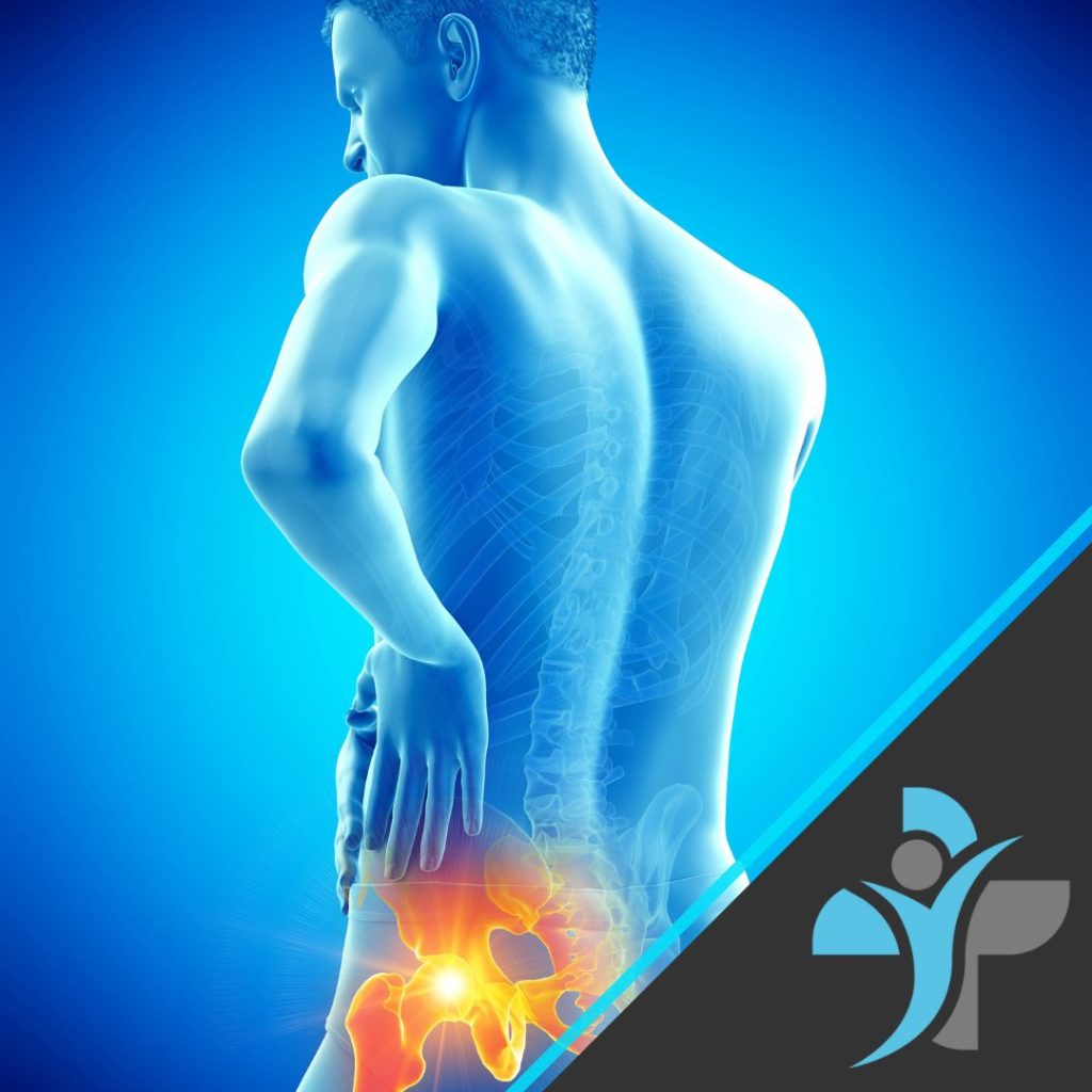
- Increased stress on the knees, hips, and lower back.
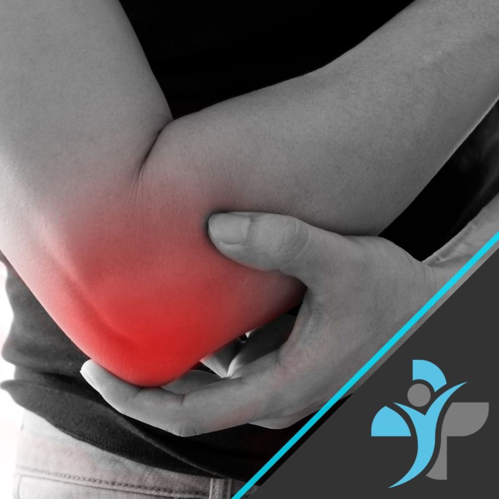
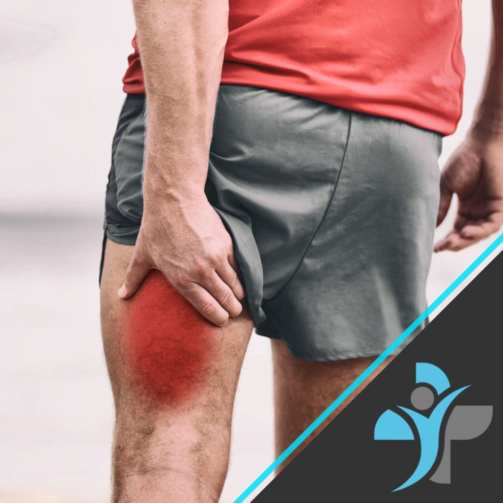
- Development of secondary conditions such as plantar fasciitis, Achilles tendonitis, metatarsalgia, or shin splints.
- Foot deformities such as flat feet, high arches, or bunions due to compensatory movements.
- Tightness in the calves upon movement.
- Nerve related issues into the foot such as tingling or numbness.
It is always best to consult a doctor or physical therapst to run through some special tests and an examination of these symptons to accurately diagnose your condition.
Diagnosis
A healthcare professional, such as a doctor, podiatrist, physical therapist, or orthopedic specialist, diagnoses equinus through:
- Physical Examination: Assessing ankle dorsiflexion range with the knee bent and extended.
- Gait Analysis: Observing walking patterns and compensations.
- Imaging Tests: X-rays, MRI, or ultrasound to check for bone or soft tissue abnormalities.
Treatment Options
Management of equinus depends on the underlying cause and severity. Common treatment approaches include:
-
Stretching and Physical Therapy:
- Calf muscle and Achilles tendon stretching exercises.
- Manual therapy to improve range of motion and flexibility.
- Massage, in particular sports massage can help greatly.
- Medical Acupuncture and or Cupping can also help. Read our article on Cupping Therapy.
-
Orthotic Devices and Footwear Modifications:
- Heel lifts or custom orthotics to accommodate limited dorsiflexion.
- Supportive footwear with a rocker sole to facilitate movement.
-
Bracing and Splinting:
- Night splints or dynamic orthotics to maintain ankle flexibility.
-
Medication and Pain Management:
- Anti-inflammatory medications to reduce pain and inflammation.
- Injections such as Botox (for spasticity-related equinus).
-
Surgical Intervention (Severe Cases):
- Gastrocnemius Recession or Achilles Tendon Lengthening to increase dorsiflexion.
- Joint Release or Bone Surgery if structural abnormalities are present.
Prognosis
With the right treatment, equinus can be management effectively and not cause any life limiting restrictions and most individuals can improve mobility and reduce pain. Early intervention is key to preventing complications and long-term musculoskeletal imbalances.
Exercises
We have provided below a short stretching program that can be used initially, but we always recommend having a bespoke plan designed based on your goals and personal circumstances.
Perform these exercises daily, ideally 2–3 times per day.
1. Standing Wall Calf Stretch
Purpose: Stretches the gastrocnemius (upper calf).
Hold for: 30–45 seconds per leg | Repeat: 2–3 times
- Stand facing a wall, placing both hands against it.
- Step one foot back while keeping it straight and pressing the heel into the floor.
- Bend the front knee slightly and lean forward.
- Keep your back leg straight and heel down.
- Switch legs and repeat.
2. Bent Knee Calf Stretch (Soleus Stretch)
Purpose: Targets the soleus (lower calf, deeper muscle).
Hold for: 30–45 seconds per leg | Repeat: 2–3 times
- Follow the same setup as the first stretch but bend the back knee slightly.
- Keep your heel on the ground as you lean into the stretch.
- You should feel the stretch lower in the calf, closer to the Achilles tendon.
3. Seated Towel Stretch
Purpose: Stretches calves and improves circulation.
Hold for: 30 seconds per leg | Repeat: 2 times
- Sit with both legs extended straight in front of you.
- Loop a towel or resistance band around the ball of one foot.
- Gently pull the towel towards you, keeping your leg straight.
- Hold and then switch sides.
4. Foam Rolling for Calves
Purpose: Releases muscle tightness and promotes blood flow.
Roll for: 1–2 minutes per leg
- Sit on the floor and place a foam roller under your calf.
- Use your hands to lift your body slightly and roll up and down from the ankle to just below the knee.
- To increase pressure, cross your other leg over the top.
5. Heel Raises (Circulation Booster)
Purpose: Activates the calf muscles and improves circulation.
Do: 15–20 reps | Repeat: 2–3 sets
- Stand with feet hip-width apart, holding onto a stable surface if needed.
- Slowly rise onto your toes, lifting your heels as high as possible.
- Hold for a second at the top, then slowly lower down.
- For an added challenge, perform single-leg heel raises.
6. Ankle Mobility Circles
Purpose: Improves circulation and prevents nerve compression.
Do: 10 circles in each direction per foot
- Sit down or lie back with your legs extended.
- Rotate your ankle in a circular motion, making big circles.
- Reverse the direction after 10 reps.
7. Nerve Glide for Sciatic and Tibial Nerves (If Numbness Persists)
Purpose: Frees up nerves that could be causing foot numbness.
Do: 10 slow reps per leg
- Sit on a chair with one leg extended.
- Point and flex your toes while sitting tall.
- You can add a head tilt (look up when flexing, down when pointing) to enhance the nerve glide.
As you can see Equinus should not be a worry or impact your life in any negative way, you should see that with positivity and some physical intervention you can live a very normal and sporty life. We treat and have treated many individuals with this condition, some of which are elite football players and it is something that can be managed effectively. If you are struggling with what you think is Equinus, then please contact our team today or arrange a booking through our online booking system to see one of our Physiotherapists or Sports Therapists.