Exercise Protocol for Back Strengthening
Back strengthening is essential for spinal health, posture, injury prevention, and functional movement. This guide explains why back training matters, who can benefit, and provides a safe, effective exercise protocol suitable for both home and gym environments.
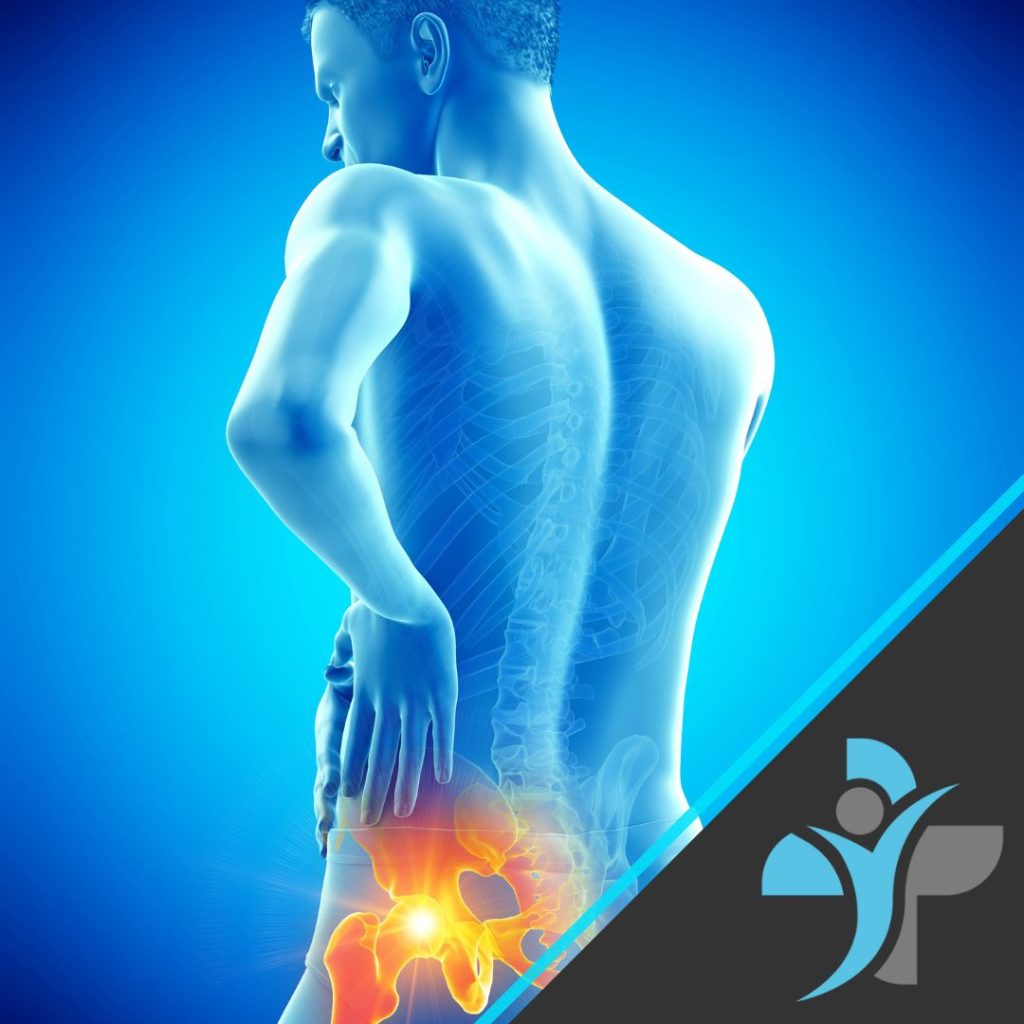
The back muscles, including the upper back, mid-back, and lower back, work closely with the core, hips, and shoulders to support everyday activities such as sitting, lifting, carrying, walking, and exercise. Weakness or poor endurance in these muscles can increase strain on the spine and contribute to pain or injury.
Why Back Training Matters?
he back muscles include the erector spinae, latissimus dorsi, rhomboids, trapezius, and deep spinal stabilisers. Together, they help to:
- Support and stabilise the spine
- Maintain upright posture
- Control movement during lifting and bending
- Transfer force between the upper and lower body
- Protect the spine during daily and sporting activities
Back weakness or poor endurance is common in individuals who:
- Sit for prolonged periods
- Have poor posture
- Perform repetitive lifting or manual work
- Experience recurrent back pain
- Lack adequate core or hip strength
Regular back strengthening may help to:
- Improve posture and spinal alignment
- Reduce the risk of back pain and recurrence
- Improve tolerance to sitting, standing, and lifting
- Enhance functional movement and daily activities
- Support sports performance and injury prevention
Who Can Benefit from Back Exercises?
Back strengthening exercises are suitable for most individuals, particularly those who:
- Experience back stiffness or weakness
- Have a history of upper or lower back pain
- Sit for long periods at work or while driving
- Perform manual handling or physically demanding work
- Want to improve posture and spinal support
Important
Always consult a qualified healthcare professional before starting a new exercise programme if you have:
- Recent or acute back injury
- Persistent or worsening back pain
- Pain radiating into the arm or leg
- Numbness, tingling, or weakness
- Diagnosed spinal conditions (e.g. disc injury, nerve compression)
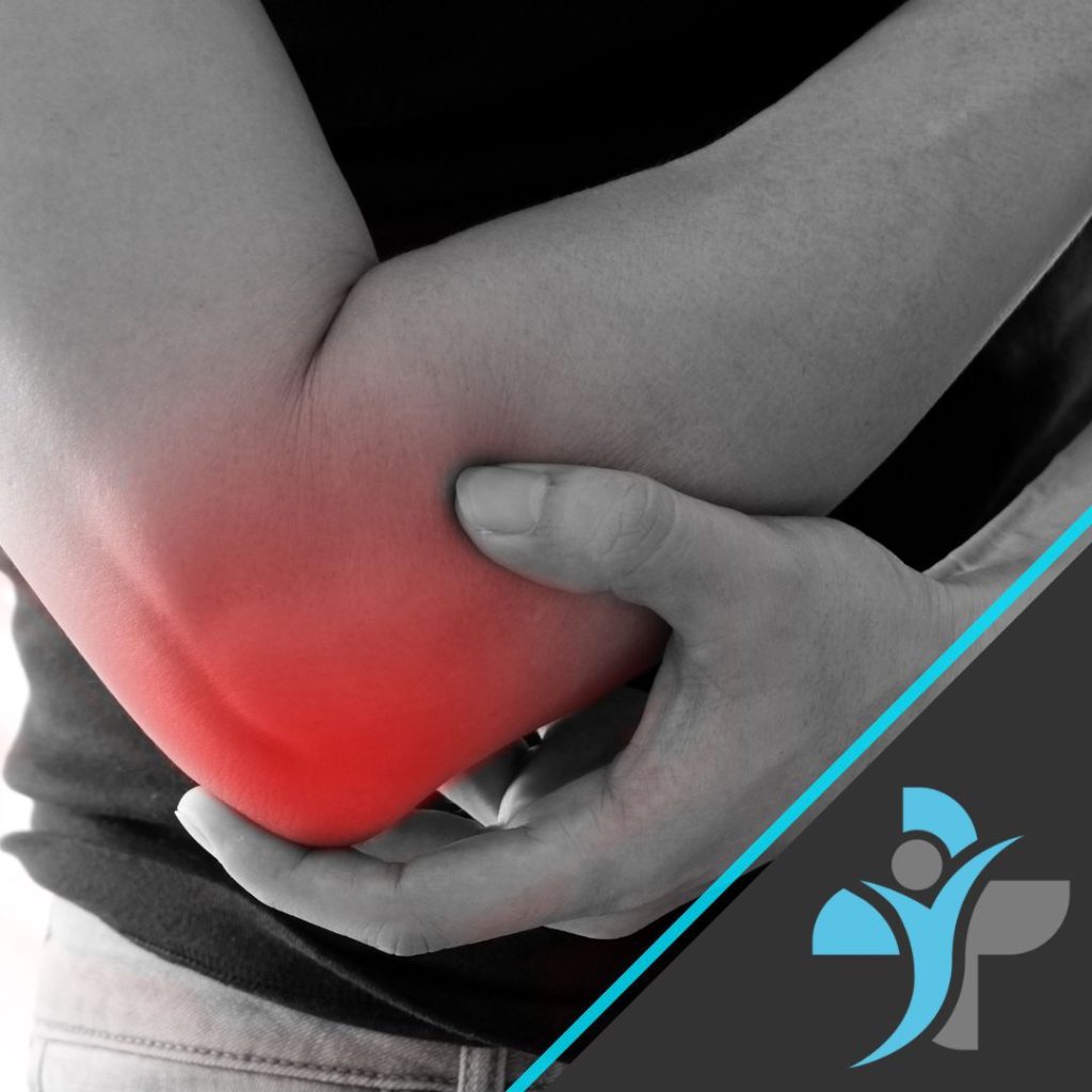
Here at Livewell Health we can help in the way of Personal Training as well as Physiotherapy and Rehabilitation of any on going injuries or issues with your back
Key Back and Postural Strengthening Exercises
-
Bird Dog
- Start on hands and knees
- Extend the opposite arm and leg
- Keep spine neutral and hips level
- Hold briefly, then switch sides
Targets: Spinal stabilisers and core control
-
Resistance Band or Cable Row
- Sit or stand tall
- Pull elbows back while squeezing shoulder blades
- Control the return
Targets: Upper and mid-back posture muscles
-
Back Extension (Floor or Bench)
- Lie prone or use a back extension bench
- Lift chest slightly while keeping neck neutral
- Avoid overextending
Targets: Lower back extensors
-
Deadlift or Hip Hinge (Bodyweight or Loaded)
- Hinge at the hips with a neutral spine
- Engage the core
- Drive through hips to stand
Targets: Posterior chain and spinal support
-
Face Pull (Band or Cable)
- Pull band or rope toward the face
- Keep elbows high
- Squeeze shoulder blades
Targets: Upper back and shoulder stabilisers
-
Wall Angels
- Stand with back against a wall
- Raise and lower arms while maintaining contact
- Keep ribs down and spine neutral
Targets: Postural endurance and upper back mobility
Safety Considerations
Back exercises should be performed with controlled movement and correct technique.
Stop exercising and seek professional advice if you experience:
- Sharp or worsening back pain
- Pain radiating into the arm or leg
- Numbness, tingling, or weakness
- Loss of bladder or bowel control (seek urgent medical care)
Avoid intense back loading during acute pain flare-ups unless guided by a qualified professional.
For guided management and physiotherapy for conditions like this, LiveWell Health’s Physiotherapy Services page includes tailored rehabilitation and hands-on treatment options.
Related Reading
For further guidance on back health, posture, and rehabilitation, these LiveWell Health resources may be helpful:
👉 Lower Back Pain – Causes, Exercises & Treatment
👉 How Poor Posture Impacts Your Spine
👉 Upper Back & Neck Pain – What You Need to Know
👉 Sports Injury Prevention & Rehabilitation
👉 Back Pain Treatment & Physiotherapy Options
Professional Support and Resources
At LiveWell Health, our multidisciplinary team provides:
- Personalised back strengthening and rehabilitation programmes
- Physiotherapy and sports therapy
- Hands-on treatment and massage therapy
- Postural assessment and movement analysis
- Education for injury prevention and long-term spinal health
If back pain, stiffness, or weakness is affecting your daily life or performance, a professional assessment can help identify the most effective treatment and exercise approach.
Book a physiotherapy or rehabilitation session
Follow us on social media for mobility tips, posture advice, and strength training updates:
Instagram: @LWHEALTH
Facebook: @livewellhealthuk